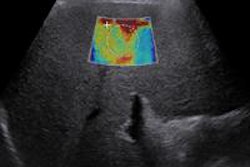
Adding shear-wave elastography (SWE) to ultrasound helps characterize suspicious breast masses, especially those in the BI-RADS 3 and BI-RADS 4 categories, according to research published online July 10 in Diagnostic and Interventional Imaging.
The findings suggest that the use of SWE could help reduce unnecessary biopsies, wrote a team led by Dr. Smriti Hari of the All India Institute of Medical Sciences in New Delhi.
"Since most [diagnostic] dilemmas arise for low-suspicion masses, the practical effect of using both techniques is for lesions near the threshold level for biopsy (i.e., BI-RADS category 3 or 4A)," the group wrote. "Some authors have shown that a large chunk (46% to 85.7%) of benign biopsies can also be reduced by adding SWE to ultrasound."
Because breast cancer tissue tends to be stiffer than healthy tissue, using SWE to assess tissue stiffness can help determine the nature of the mass, Hari and colleagues wrote. Masses categorized as BI-RADS 3 are recommended for short-term follow-up, while those categorized at BI-RADS 4 can be trickier, with malignancy rates ranging from 2% to 94%.
"BI-RADS category 4 masses are considered suspicious, and the diagnosis is often challenging," they wrote. "[It] would be worthwhile to avoid delay in diagnosis of even a few malignancies among these lesions."
Hari's team included data from 119 women with a single breast mass between June 2013 and November 2014. All of the women underwent a clinical breast exam, ultrasound, SWE, and ultrasound-guided core biopsy of the breast mass; the masses were assigned a BI-RADS category. The researchers estimated cutoff values for differentiating benign and malignant masses on SWE, taking elasticity parameters (mean, minimum, and maximum) and color into account.
BI-RADS 3 and 4A (low suspicion) masses were either upgraded or downgraded by combining SWE and ultrasound findings. Finally, the group compared the sensitivity, specificity, and positive and negative predictive values of modified BI-RADS categories (which included both SWE and ultrasound data) and BI-RADS categories established by ultrasound alone.
On histopathology, 57 of 119 breast masses (48%) were benign and 62 (52%) were malignant. Ultrasound alone had a sensitivity rate of 96.8% and a specificity rate of 70.2%. However, adding SWE to ultrasound improved the specificity of BI-RADS categories by particular characteristics, specifically by color and elasticity maximum and mean values.
| B-mode ultrasound alone vs. B-mode + SWE for characterizing breast masses | ||||
| Measure | B-mode ultrasound alone | B-mode ultrasound plus SWE color | B-mode ultrasound plus SWE maximum elasticity | B-mode ultrasound plus SWE mean elasticity |
| Sensitivity | 96.8% | 96.8% | 96.8% | 96.8% |
| Specificity | 70.2% | 78.9% | 75.4% | 75.4% |
| Positive predictive value | 77.9% | 83.3% | 81.1% | 81.1% |
| Negative predictive value | 95.2% | 95.7% | 95.6% | 95.6% |
SWE is a useful technique for characterizing breast masses, Hari and colleagues concluded.
"[Elasticity mean and maximum] are the best SWE parameters for characterizing breast masses," they wrote. "SWE, used in addition to ultrasound, improves characterization of BI-RADS 3 and 4A masses."