Despite little growth in the prevalence of peripheral arterial disease (PAD), utilization of tests for the condition grew rapidly from 2001 to 2013. This increase may reflect overutilization and possibly the effects of self-referral among cardiologists and surgeons, according to a new study.
On the bright side, utilization rates have flattened since 2010 for duplex ultrasound of lower extremity arteries and decreased for noninvasive physiologic testing of extremity arteries (NPT), according to a review of Medicare data files by researchers from Thomas Jefferson University. Radiologists have lowered their ultrasound utilization rate; however, surgeons and cardiologists have continued to show an increase, raising the question of a possible self-referral problem, according to the team led by Dr. David C. Levin.
Levin presented the results at the recent RSNA 2015 meeting in Chicago.
PAD screening controversy
Unlike coronary artery disease or carotid artery disease, which can result in heart attack or stroke, PAD only rarely results in catastrophic consequences if not detected early, Levin said.
"There are almost no catastrophes [from PAD]; it is kind of a disease that has a very gradual onset," he said. "If you're late in diagnosing it, it probably doesn't make that much difference to the clinical outcome of the patient. You don't really need to treat peripheral arterial disease until it's really quite symptomatic. Testing for the early presence of disease is not urgent."
At the same time, the prevalence of PAD is increasing at a very slow rate of 1% per year in developed countries, according to a recent study. Nonetheless, screening for PAD remains a controversial issue. The Society of Interventional Radiology recommends screening, while the Society for Vascular Surgery is against it, Levin said.
The researchers set out to study recent utilization trends for the two most common noninvasive tests used to screen for and initially diagnose PAD: duplex ultrasound of the lower extremities and noninvasive physiologic testing, which includes procedures such as the ankle-brachial index, pulse volume recordings, segmental pressure measurements, and bidirectional Doppler waveforms.
The team accessed Medicare Part B Physician/Supplier Procedure Summary Master Files for 2001-2013, which provide data such as exam volume and allowed payments for each CPT code. Utilization rates per 100,000 Medicare beneficiaries were calculated for two duplex ultrasound CPT codes (93925 and 93926) and three NPT CPT codes (93922, 93923, and 93924). The researchers then used the Medicare specialty codes to identify the physician specialties for the studies.
Increase in utilization
In bad news, the group found a significant increase in exam volume and utilization rates per 1,000 Medicare beneficiaries for both duplex ultrasound and NPT studies.
 Images courtesy of Dr. David C. Levin.
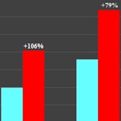
The utilization rates per 1,000 Medicare beneficiaries have increased by 88% for ultrasound and by 63% for NPT from 2001 to 2013.
"At a time when the prevalence of PAD was growing very slowly, the use of these tests grew rapidly," he said. "This raises a concern about overutilization."
| Utilization of extremity arterial ultrasound for PAD by specialty | ||
| Specialty | 2013 | Increase from 2001 |
| Radiology | 210,477 | 93% |
| Surgery | 258,104 | 108% |
| Cardiology | 187,275 | 267% |
On the bright side, growth in ultrasound utilization flattened out and NPT use has declined since 2010. This trend is likely due to overall efforts in healthcare and imaging to restrict rapid growth in testing, Levin said.
Surgeons, cardiologists buck trend
The researchers also found that radiologists' use of ultrasound has been flat since around 2009, while surgeons and cardiologists continued to perform more studies. That raises concern over self-referral issues, Levin said.
The team did find a decline in NPT among all specialties since 2010.
Levin noted that the increases in the overall utilization rates of duplex ultrasound and NPT were not related to an increase in screening, as Medicare does not cover the use of these tests for screening purposes.
It's good, though, that radiologists retain a strong role in PAD testing, especially in lower extremity duplex ultrasound, Levin said.
"Angiography and peripheral vascular interventions have been taken over pretty much by surgeons, and I think that's unfortunate," he told AuntMinnie.com. "I don't think they're nearly as well-trained as radiologists and their results are not as good. That was a development I was really unhappy about."