Dear AuntMinnie Member,
Are too few female medical students choosing to become radiologists? That was the question asked this week during the American College of Radiology's ACR 2015 meeting in Washington, DC.
Researchers from Rutgers University presented their study on whether the proportion of female radiologists had changed much compared to 10 years ago, when the disparity was first recognized.
But while the discipline has made progress in some areas, the number of women who pick careers in radiology has remained stuck at about 27%, even as the percentage of female medical students stands at 46%. Learn more by clicking here.
How good is bedside US in the ER?
In other news from ACR 2015, a research group from California is raising questions about the effectiveness of point-of-care ultrasound for first-trimester obstetric patients in the emergency room (ER) setting.
Such scans are becoming ubiquitous as ER physicians take advantage of ultrasound's unique benefits for scanning obstetric patients. However, ultrasound is an operator-dependent modality that can be challenging to interpret -- so should ER patients also be sent on for radiology ultrasound exams?
The researchers found that it might be a good idea. Point-of-care ultrasound studies had a 24% discrepancy rate with radiology ultrasound scans, indicating that ER physicians might be missing some pathology. Get the rest of the story by clicking here.
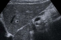
In other ultrasound news, another group from California found that many scans for the surveillance of hepatocellular carcinoma may not be good enough due to problems with visualization and penetration of the liver. That article can be reached by clicking here.
Get these stories and more in our Ultrasound Community, at ultrasound.auntminnie.com.







![Examples of ultrasound findings and techniques. (A) Images in a 39-year-old male patient with a mass in the left thigh. The mass is heterogeneous on the B-mode US image (compared with the patient in D) and showed increased microvascularity (superb microvascular imaging [SMI]) and shear-wave elastography (SWE) values. Undifferentiated pleomorphic sarcoma was diagnosed at biopsy (with pleomorphic rhabdomyosarcoma in surgical specimen). (B) Images in an 18-year-old male patient with a mass in the left leg. The mass is hypoechoic on the B-mode image, with no other findings suggestive of malignancy. The lesion is in contact with the cortex of the tibia, which is slightly irregular. CT revealed a doubtful anteromedial tibial erosion. The microvascular study demonstrated high vascularization, suggestive of malignancy. Periosteal Ewing sarcoma was diagnosed with both histologic and immunohistochemical confirmation. (C) Images in a 69-year-old female patient with a lump growing on the outside of the left leg. Multiple SWE examinations were performed (please note the high values obtained in the measurements, whereas the color map highlights the stiffness relative to adjacent tissues). SMI showed areas of increased vascularization to target for sampling. Undifferentiated spindle cell sarcoma was diagnosed at biopsy, with residual leiomyosarcoma in the surgical specimen after neoadjuvant therapy. (D) Images in a 56-year-old female patient with a mass in the right thigh. The mass is heterogeneous at both B-mode ultrasound (similar to patient A) and MRI (coronal T2-weighted spectral attenuated inversion recovery [SPAIR]; T1-weighted pre-contrast and postcontrast imaging), which even shows uptake after the administration of paramagnetic contrast material, which is traditionally suggestive of malignancy. Low values at SMI and elastography are suggestive of benignity. Spindle cell lipoma was diagnosed at biopsy, with atypical spindle cell lipomatous tumor in the surgical specimen.](https://img.auntminnie.com/mindful/smg/workspaces/default/uploads/2026/08/images-radiol250278fig2.APCFLSvX6p.jpg?auto=format%2Ccompress&fit=crop&h=112&q=70&w=112)


