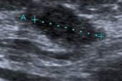
Breast ultrasound findings can obviate the need for excisional biopsy in most cases of teenage girls with breast lumps, according to recent research from Loyola University Health System.
In an article published in the Journal of Ultrasound in Medicine (April 2015, Vol. 34:4, pp. 617-625), Loyola researchers shared their research project that included 37 teenage girls with a total of 45 breast lumps. The study sought to determine whether the breast mass size at an initial ultrasound and growth at follow-up ultrasound could be used to determine if a lump could be managed conservatively or if a tissue biopsy was required.
The 37 girls were divided into three study groups. The first group (nine masses) received a follow-up ultrasound and a biopsy, while a second group (13 masses) only received a biopsy. The third group (23 masses) received a follow-up ultrasound without a biopsy. None of the lumps showed changes in appearance at follow-up ultrasound, and all the lumps that were biopsied were benign, according to the researchers.
In other analyses of lump dimension, volume, and volume change, the researchers concluded that biopsies could have been reduced in 89% of girls in the first study group and 96% in the third group if biopsies were only performed on breast masses with a largest dimension greater than 3 cm and a volume change per month greater than 16%.
Lead author Dr. Aruna Vade and colleagues recommend that excisional biopsy be performed only on solid breast masses that are sonographically suspicious for malignancy or breast masses that show progressive growth or are found in patients who have a known primary malignant tumor or family history of cancer.








![Examples of ultrasound findings and techniques. (A) Images in a 39-year-old male patient with a mass in the left thigh. The mass is heterogeneous on the B-mode US image (compared with the patient in D) and showed increased microvascularity (superb microvascular imaging [SMI]) and shear-wave elastography (SWE) values. Undifferentiated pleomorphic sarcoma was diagnosed at biopsy (with pleomorphic rhabdomyosarcoma in surgical specimen). (B) Images in an 18-year-old male patient with a mass in the left leg. The mass is hypoechoic on the B-mode image, with no other findings suggestive of malignancy. The lesion is in contact with the cortex of the tibia, which is slightly irregular. CT revealed a doubtful anteromedial tibial erosion. The microvascular study demonstrated high vascularization, suggestive of malignancy. Periosteal Ewing sarcoma was diagnosed with both histologic and immunohistochemical confirmation. (C) Images in a 69-year-old female patient with a lump growing on the outside of the left leg. Multiple SWE examinations were performed (please note the high values obtained in the measurements, whereas the color map highlights the stiffness relative to adjacent tissues). SMI showed areas of increased vascularization to target for sampling. Undifferentiated spindle cell sarcoma was diagnosed at biopsy, with residual leiomyosarcoma in the surgical specimen after neoadjuvant therapy. (D) Images in a 56-year-old female patient with a mass in the right thigh. The mass is heterogeneous at both B-mode ultrasound (similar to patient A) and MRI (coronal T2-weighted spectral attenuated inversion recovery [SPAIR]; T1-weighted pre-contrast and postcontrast imaging), which even shows uptake after the administration of paramagnetic contrast material, which is traditionally suggestive of malignancy. Low values at SMI and elastography are suggestive of benignity. Spindle cell lipoma was diagnosed at biopsy, with atypical spindle cell lipomatous tumor in the surgical specimen.](https://img.auntminnie.com/mindful/smg/workspaces/default/uploads/2026/08/images-radiol250278fig2.APCFLSvX6p.jpg?auto=format%2Ccompress&fit=crop&h=112&q=70&w=112)


