Advocacy group Public Citizen is criticizing health screenings offered by HealthFair Health Screening, a company that offers mobile screening services.
In a June 19 press release, Public Citizen urged 20 hospitals in eight states to cut ties with HealthFair, calling the company's cardiovascular screening programs "unethical" and "much more likely to do harm than good."
HealthFair offers a variety of health screening tests, some of which use medical imaging technologies, such as abdominal aortic aneurysm screening with ultrasound, carotid artery ultrasound, and bone density ultrasound.
Public Citizen claims that HealthFair partners with hospitals and medical institutions to offer screening packages to individuals living in their geographic areas. The packages are marketed by both HealthFair and the hospitals using tactics that Public Citizen described as "fearmongering."
Public Citizen went on to charge that HealthFair pitches the tests on a widespread basis "without identifying who has relevant risk factors that would make each of the screening tests medically appropriate." In a letter to the hospitals, Public Citizen claims that no evidence-based guidelines issued by major medical associations support the use of six of the tests for widespread screening of asymptomatic people. A sample of the Public Citizen letter is available here.
Such "indiscriminate testing" is more likely to lead to harm than good, according to the group, due to false-positive results that will require additional tests and treatments that could cause harm and patient anxiety.
Public Citizen sent the letters to hospitals in Arizona, California, Florida, Illinois, Indiana, Iowa, Ohio, and Virginia.






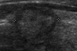



![Examples of ultrasound findings and techniques. (A) Images in a 39-year-old male patient with a mass in the left thigh. The mass is heterogeneous on the B-mode US image (compared with the patient in D) and showed increased microvascularity (superb microvascular imaging [SMI]) and shear-wave elastography (SWE) values. Undifferentiated pleomorphic sarcoma was diagnosed at biopsy (with pleomorphic rhabdomyosarcoma in surgical specimen). (B) Images in an 18-year-old male patient with a mass in the left leg. The mass is hypoechoic on the B-mode image, with no other findings suggestive of malignancy. The lesion is in contact with the cortex of the tibia, which is slightly irregular. CT revealed a doubtful anteromedial tibial erosion. The microvascular study demonstrated high vascularization, suggestive of malignancy. Periosteal Ewing sarcoma was diagnosed with both histologic and immunohistochemical confirmation. (C) Images in a 69-year-old female patient with a lump growing on the outside of the left leg. Multiple SWE examinations were performed (please note the high values obtained in the measurements, whereas the color map highlights the stiffness relative to adjacent tissues). SMI showed areas of increased vascularization to target for sampling. Undifferentiated spindle cell sarcoma was diagnosed at biopsy, with residual leiomyosarcoma in the surgical specimen after neoadjuvant therapy. (D) Images in a 56-year-old female patient with a mass in the right thigh. The mass is heterogeneous at both B-mode ultrasound (similar to patient A) and MRI (coronal T2-weighted spectral attenuated inversion recovery [SPAIR]; T1-weighted pre-contrast and postcontrast imaging), which even shows uptake after the administration of paramagnetic contrast material, which is traditionally suggestive of malignancy. Low values at SMI and elastography are suggestive of benignity. Spindle cell lipoma was diagnosed at biopsy, with atypical spindle cell lipomatous tumor in the surgical specimen.](https://img.auntminnie.com/mindful/smg/workspaces/default/uploads/2026/08/images-radiol250278fig2.APCFLSvX6p.jpg?auto=format%2Ccompress&dpr=2&fit=crop&h=167&q=70&w=250)
