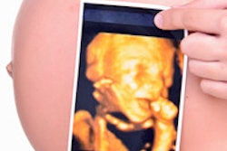
An investigative report has caused a stir in Canada after it revealed that keepsake ultrasound clinics are willing to provide the gender of fetuses before 20 weeks of gestation. This is early enough for women to request an abortion and feeds into the phenomenon of female feticide in some immigrant communities in Canada.
In a June 12 broadcast, reporters with the Canadian Broadcasting Corporation (CBC) used hidden cameras and documented that of 22 keepsake 3D ultrasound clinics visited, 15 agreed to provide an ultrasound that would reveal the gender of the fetus before 20 weeks of pregnancy. Five of the facilities agreed to provide gender testing as early as 14 weeks of gestation. The original report can be reached by clicking here.
Keepsake ultrasound sites have blossomed across Canada over the past 10 years. In contrast, some Canadian hospitals located in close proximity to large South Asian communities, including several in Ontario, have prohibited gender notification due to its relationship to the abortion of female fetuses.
The broadcast of the undercover investigation led Dr. Michiel Van den Hof, a spokesperson for the Society of Obstetricians and Gynaecologists of Canada (SOGC), to tell the CBC that his society backs a complete ban on keepsake ultrasounds.
When AuntMinnie.com contacted SOGC for a comment, spokesperson Natalie Wright pointed to an April 2007 SOGC policy statement that recommended a "complete ban on the nonmedical use of fetal ultrasound." Wright said that although the "document was published five years ago, its content accurately reflects the SOGC's position today."
However, the SOGC has also said in the past that it supports a patient's right to know the gender of the child.
A paper published this week documents a significantly higher male-to-female ratio among firstborn children to women living in Ontario who came from South Korea and India. The male-to-female ratio was even higher for Indian-born women who had more than one child (Canadian Medical Association Journal, June 12, 2012, Vol. 184, pp. E492-E496).
"Our findings raise the possibility that couples originating from India may be more likely than Canadian-born couples to use prenatal sex determination and terminate a second or subsequent pregnancy if the fetus is female," wrote Dr. Joel Ray, from St. Michael's Hospital in Toronto, and co-authors.
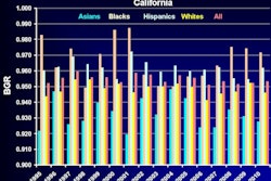
U.S. researchers have discovered similar trends among Asian immigrant cultures in California, a phenomenon that also has been tied to keepsake ultrasound centers.
When asked by AuntMinnie.com about the best way to combat this phenomenon, Ray said, "I do not think that a federal law or SOGC mandate will fully [dissuade] determined women going to other countries to get a fetal sex ultrasound or pregnancy termination because of fetal sex."
"Rather, we need to emphasize the value of females -- born and unborn -- and to stress this through community-specific endeavors," he said.