Automated breast ultrasound (ABUS) finds cancers in dense breast tissue that mammography alone would not, according to a recent study. The findings indicate that ABUS could have a role in a screening environment for women with dense breasts.
Ultrasound has demonstrated its value as an adjunct to conventional mammography for screening women, particularly those with dense breast tissue, said Dr. Judy Dean, a radiologist in private practice in Santa Barbara, CA. However, radiologists are concerned that using the technology manually for screening will take too much time.
"Numerous studies beginning in the 1990s have shown the value of ultrasound in detecting cancers not seen on mammography in women with dense breast tissue, but radiologists have not embraced the modality because of the time and training it takes to perform the examinations," Dean told attendees at a session at the RSNA 2011 meeting. "[But we found that] cancer detections increased by 63% with [ABUS] after radiologists received only four hours of training."
Automated breast ultrasound acquires overlapping scan rows in an axial plane, Dean said. The images are reviewed by the radiologist as a cine loop, and cancers are identified by the disruption of structures. Spatial registration allows for localization of identified findings.
For the current study, Dean and colleagues examined asymptomatic women having routine mammography. The study included 3,778 digital mammograms and ABUS studies; all women had dense breast tissue (BI-RADS 3 or 4 density score) or a high risk of breast cancer. Diagnostic yield and specificity of mammography were compared to results with ABUS and the two modalities combined. The study was blinded but not randomized, she said.
All imaging studies were performed by technical staff and interpreted later by a radiologist, Dean told session attendees. The exams were handled as screening studies, with recall for workup of suspicious findings.
Mammography alone found 4.5 nonpalpable cancers per 1,000 women; adding ABUS increased the number to 7.9 per 1,000, an increase in yield of 76%. ABUS found 13 additional cancers, all of which were invasive, Dean said. The average size of the invasive cancers found was 14 mm with digital mammography and 9 mm with ABUS.
There were no significant changes in specificity and positive predictive value (PPV) between the two modalities. Mammography's specificity was 99.5% and its PPV was 50%; the specificity of ABUS was 99.4% and its PPV was 43.6%. The combined specificity of the modalities was 99%, while their combined PPV was 44.1%, according to Dean.
![]() |
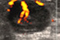
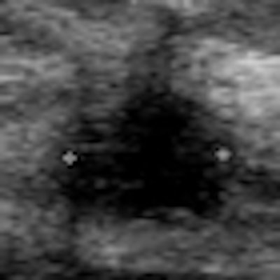
| Mediolateral oblique (MLO) views of a 56-year-old woman whose sister had breast cancer are normal (above). But automated breast ultrasound (below) showed a small mass in the left breast. This image is from a focused breast ultrasound exam, performed because of the finding detected on automated ultrasound. Biopsy confirmed a 10-mm invasive ductal carcinoma. All images courtesy of Dr. Judy Dean. |
![]() |
While early detection of breast cancer with mammography can prevent breast cancer deaths, ABUS can help find those cancers mammography might miss in high-risk or dense-breasted women, Dean concluded.
| Dean is a stockholder in SonoCine and iCAD, and she is also a consultant to and reader study participant for U-Systems. |





![Examples of ultrasound findings and techniques. (A) Images in a 39-year-old male patient with a mass in the left thigh. The mass is heterogeneous on the B-mode US image (compared with the patient in D) and showed increased microvascularity (superb microvascular imaging [SMI]) and shear-wave elastography (SWE) values. Undifferentiated pleomorphic sarcoma was diagnosed at biopsy (with pleomorphic rhabdomyosarcoma in surgical specimen). (B) Images in an 18-year-old male patient with a mass in the left leg. The mass is hypoechoic on the B-mode image, with no other findings suggestive of malignancy. The lesion is in contact with the cortex of the tibia, which is slightly irregular. CT revealed a doubtful anteromedial tibial erosion. The microvascular study demonstrated high vascularization, suggestive of malignancy. Periosteal Ewing sarcoma was diagnosed with both histologic and immunohistochemical confirmation. (C) Images in a 69-year-old female patient with a lump growing on the outside of the left leg. Multiple SWE examinations were performed (please note the high values obtained in the measurements, whereas the color map highlights the stiffness relative to adjacent tissues). SMI showed areas of increased vascularization to target for sampling. Undifferentiated spindle cell sarcoma was diagnosed at biopsy, with residual leiomyosarcoma in the surgical specimen after neoadjuvant therapy. (D) Images in a 56-year-old female patient with a mass in the right thigh. The mass is heterogeneous at both B-mode ultrasound (similar to patient A) and MRI (coronal T2-weighted spectral attenuated inversion recovery [SPAIR]; T1-weighted pre-contrast and postcontrast imaging), which even shows uptake after the administration of paramagnetic contrast material, which is traditionally suggestive of malignancy. Low values at SMI and elastography are suggestive of benignity. Spindle cell lipoma was diagnosed at biopsy, with atypical spindle cell lipomatous tumor in the surgical specimen.](https://img.auntminnie.com/mindful/smg/workspaces/default/uploads/2026/08/images-radiol250278fig2.APCFLSvX6p.jpg?auto=format%2Ccompress&fit=crop&h=100&q=70&w=100)


![Examples of ultrasound findings and techniques. (A) Images in a 39-year-old male patient with a mass in the left thigh. The mass is heterogeneous on the B-mode US image (compared with the patient in D) and showed increased microvascularity (superb microvascular imaging [SMI]) and shear-wave elastography (SWE) values. Undifferentiated pleomorphic sarcoma was diagnosed at biopsy (with pleomorphic rhabdomyosarcoma in surgical specimen). (B) Images in an 18-year-old male patient with a mass in the left leg. The mass is hypoechoic on the B-mode image, with no other findings suggestive of malignancy. The lesion is in contact with the cortex of the tibia, which is slightly irregular. CT revealed a doubtful anteromedial tibial erosion. The microvascular study demonstrated high vascularization, suggestive of malignancy. Periosteal Ewing sarcoma was diagnosed with both histologic and immunohistochemical confirmation. (C) Images in a 69-year-old female patient with a lump growing on the outside of the left leg. Multiple SWE examinations were performed (please note the high values obtained in the measurements, whereas the color map highlights the stiffness relative to adjacent tissues). SMI showed areas of increased vascularization to target for sampling. Undifferentiated spindle cell sarcoma was diagnosed at biopsy, with residual leiomyosarcoma in the surgical specimen after neoadjuvant therapy. (D) Images in a 56-year-old female patient with a mass in the right thigh. The mass is heterogeneous at both B-mode ultrasound (similar to patient A) and MRI (coronal T2-weighted spectral attenuated inversion recovery [SPAIR]; T1-weighted pre-contrast and postcontrast imaging), which even shows uptake after the administration of paramagnetic contrast material, which is traditionally suggestive of malignancy. Low values at SMI and elastography are suggestive of benignity. Spindle cell lipoma was diagnosed at biopsy, with atypical spindle cell lipomatous tumor in the surgical specimen.](https://img.auntminnie.com/mindful/smg/workspaces/default/uploads/2026/08/images-radiol250278fig2.APCFLSvX6p.jpg?auto=format%2Ccompress&fit=crop&h=112&q=70&w=112)