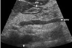
(Ultrasound Review) A recent study published in American Journal of Roentgenology compared real-time spatial compound sonography with conventional musculoskeletal (MSK) ultrasound imaging.
According to radiologists at Thomas Jefferson University Hospital in Philadelphia, "real-time spatial compound sonography significantly improved sonographic image quality in the musculoskeletal system when compared with conventional high-resolution sonography."
Spatial compound sonography is a relatively new technique that uses multiple, differing angles of insonation to create an ultrasound image. This form of image compounding reduces image noise and speckle, and increases tissue plane definition.
"As the image is generated from more angles of insonation, the likelihood is greater that one of these angles will be perpendicular to specular reflectors, generate a higher echo amplitude, and thereby reduce anisotropic effects," they said.
Fifty-nine image pairs were digitally stored and reviewed by sonologists. Images of both normal anatomy and pathology using conventional ultrasound and compounded imaging were compared, and an assessment of image quality was made.
"Each image was rated for definition of tissue planes, amount of speckle and other noise, and image detail using a 5-point scale: 1, unacceptable; 2, poor quality but marginally acceptable; 3, fair quality and acceptable; 4, good quality; and 5, excellent or outstanding quality," they reported.
According to the results, "an analysis of variance revealed that real-time spatial compound sonography significantly improved definition of soft-tissue planes, reduced speckle and other noise, and improved image detail when compared with conventional high-resolution sonography."
Compound imaging requires more lines of sight resulting in lower frame rates and reduced temporal resolution. In MSK imaging, the structures being imaged were still and superficial, and so did not cause significant image degradation. When dynamic imaging is performed they suggested that higher frame rates should be selected.
They concluded, "spatial compound sonography reduces image artifacts and thereby improves tissue-plane definition. Because musculoskeletal sonography is highly dependent on image quality and tissue-plane definition, real-time spatial compound sonography represents an important development in the field."
Advantages of real-time spatial compound sonography of the musculoskeletal system versus conventional sonographyLin, D et al
Division of ultrasound, department of radiology, Thomas Jefferson University Hospital, Philadelphia, PA
AJR 2002 December; 179:1629-1631
By Ultrasound Review
December 17, 2002
Copyright © 2002 AuntMinnie.com

![Examples of ultrasound findings and techniques. (A) Images in a 39-year-old male patient with a mass in the left thigh. The mass is heterogeneous on the B-mode US image (compared with the patient in D) and showed increased microvascularity (superb microvascular imaging [SMI]) and shear-wave elastography (SWE) values. Undifferentiated pleomorphic sarcoma was diagnosed at biopsy (with pleomorphic rhabdomyosarcoma in surgical specimen). (B) Images in an 18-year-old male patient with a mass in the left leg. The mass is hypoechoic on the B-mode image, with no other findings suggestive of malignancy. The lesion is in contact with the cortex of the tibia, which is slightly irregular. CT revealed a doubtful anteromedial tibial erosion. The microvascular study demonstrated high vascularization, suggestive of malignancy. Periosteal Ewing sarcoma was diagnosed with both histologic and immunohistochemical confirmation. (C) Images in a 69-year-old female patient with a lump growing on the outside of the left leg. Multiple SWE examinations were performed (please note the high values obtained in the measurements, whereas the color map highlights the stiffness relative to adjacent tissues). SMI showed areas of increased vascularization to target for sampling. Undifferentiated spindle cell sarcoma was diagnosed at biopsy, with residual leiomyosarcoma in the surgical specimen after neoadjuvant therapy. (D) Images in a 56-year-old female patient with a mass in the right thigh. The mass is heterogeneous at both B-mode ultrasound (similar to patient A) and MRI (coronal T2-weighted spectral attenuated inversion recovery [SPAIR]; T1-weighted pre-contrast and postcontrast imaging), which even shows uptake after the administration of paramagnetic contrast material, which is traditionally suggestive of malignancy. Low values at SMI and elastography are suggestive of benignity. Spindle cell lipoma was diagnosed at biopsy, with atypical spindle cell lipomatous tumor in the surgical specimen.](https://img.auntminnie.com/mindful/smg/workspaces/default/uploads/2026/08/images-radiol250278fig2.APCFLSvX6p.jpg?auto=format%2Ccompress&fit=crop&h=100&q=70&w=100)


![Examples of ultrasound findings and techniques. (A) Images in a 39-year-old male patient with a mass in the left thigh. The mass is heterogeneous on the B-mode US image (compared with the patient in D) and showed increased microvascularity (superb microvascular imaging [SMI]) and shear-wave elastography (SWE) values. Undifferentiated pleomorphic sarcoma was diagnosed at biopsy (with pleomorphic rhabdomyosarcoma in surgical specimen). (B) Images in an 18-year-old male patient with a mass in the left leg. The mass is hypoechoic on the B-mode image, with no other findings suggestive of malignancy. The lesion is in contact with the cortex of the tibia, which is slightly irregular. CT revealed a doubtful anteromedial tibial erosion. The microvascular study demonstrated high vascularization, suggestive of malignancy. Periosteal Ewing sarcoma was diagnosed with both histologic and immunohistochemical confirmation. (C) Images in a 69-year-old female patient with a lump growing on the outside of the left leg. Multiple SWE examinations were performed (please note the high values obtained in the measurements, whereas the color map highlights the stiffness relative to adjacent tissues). SMI showed areas of increased vascularization to target for sampling. Undifferentiated spindle cell sarcoma was diagnosed at biopsy, with residual leiomyosarcoma in the surgical specimen after neoadjuvant therapy. (D) Images in a 56-year-old female patient with a mass in the right thigh. The mass is heterogeneous at both B-mode ultrasound (similar to patient A) and MRI (coronal T2-weighted spectral attenuated inversion recovery [SPAIR]; T1-weighted pre-contrast and postcontrast imaging), which even shows uptake after the administration of paramagnetic contrast material, which is traditionally suggestive of malignancy. Low values at SMI and elastography are suggestive of benignity. Spindle cell lipoma was diagnosed at biopsy, with atypical spindle cell lipomatous tumor in the surgical specimen.](https://img.auntminnie.com/mindful/smg/workspaces/default/uploads/2026/08/images-radiol250278fig2.APCFLSvX6p.jpg?auto=format%2Ccompress&fit=crop&h=112&q=70&w=112)