(Ultrasound Review) According to researchers at the University of California San Diego Medical Center, a diagnosis of marginal placental cord insertion (PCI) is not associated with growth retardation or preterm delivery.
"Abnormalities of placental cord insertion have been linked to a variety of poor obstetric outcomes," they reported. In particular, velamentous PCI has an association with poor fetal growth, fetal anomalies, preterm delivery and pregnancy complications, the authors noted.
"This study was designed to evaluate the impact of prenatally diagnosed marginal PCI on birth weight and duration of pregnancy," they wrote in the Journal of Ultrasound in Medicine.
Radiologists and perinatologists combined to retrospectively study 100 singleton pregnancies that were found to have a cord insertion 2cm or less from any placental edge. The PCI was routinely identified during the second trimester morphology scan. The relationship of PCI to adjacent placental edges and cord vessels as they entered the placenta was visualized. In addition, chorionic vessels were identified to avoid confusion with cord vessels.
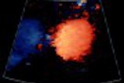
In order to maximize sonographic specificity, a combination of color and power Doppler and grayscale was used to identify the PCI. Any low-lying, bipartite or succinturiate lobed placentas were scrutinized using transvaginal scanning to check for vasa previa.
The authors asserted that the PCI could be identified in all fetuses during the second trimester. Results demonstrated no significant association between marginal PCI and fetal growth restriction or premature delivery. Because of the retrospective nature of this study, the progression of marginal PCI to velamentous insertion was not evaluated. The authors recommend using a cutoff of 0.5cm from any adjacent placental edge for velamentous PCI and suggested that these pregnancies be carefully monitored sonographically for fetal growth.
Sonographic prenatal diagnosis of marginal placental cord insertionCC Liu et al
Department of radiology, University of California San Diego Medical, San Diego, CA
J Ultrasound Med 2002 June; 21: 627-632
By Ultrasound Review
July 24, 2002
Copyright © 2002 AuntMinnie.com
![Examples of ultrasound findings and techniques. (A) Images in a 39-year-old male patient with a mass in the left thigh. The mass is heterogeneous on the B-mode US image (compared with the patient in D) and showed increased microvascularity (superb microvascular imaging [SMI]) and shear-wave elastography (SWE) values. Undifferentiated pleomorphic sarcoma was diagnosed at biopsy (with pleomorphic rhabdomyosarcoma in surgical specimen). (B) Images in an 18-year-old male patient with a mass in the left leg. The mass is hypoechoic on the B-mode image, with no other findings suggestive of malignancy. The lesion is in contact with the cortex of the tibia, which is slightly irregular. CT revealed a doubtful anteromedial tibial erosion. The microvascular study demonstrated high vascularization, suggestive of malignancy. Periosteal Ewing sarcoma was diagnosed with both histologic and immunohistochemical confirmation. (C) Images in a 69-year-old female patient with a lump growing on the outside of the left leg. Multiple SWE examinations were performed (please note the high values obtained in the measurements, whereas the color map highlights the stiffness relative to adjacent tissues). SMI showed areas of increased vascularization to target for sampling. Undifferentiated spindle cell sarcoma was diagnosed at biopsy, with residual leiomyosarcoma in the surgical specimen after neoadjuvant therapy. (D) Images in a 56-year-old female patient with a mass in the right thigh. The mass is heterogeneous at both B-mode ultrasound (similar to patient A) and MRI (coronal T2-weighted spectral attenuated inversion recovery [SPAIR]; T1-weighted pre-contrast and postcontrast imaging), which even shows uptake after the administration of paramagnetic contrast material, which is traditionally suggestive of malignancy. Low values at SMI and elastography are suggestive of benignity. Spindle cell lipoma was diagnosed at biopsy, with atypical spindle cell lipomatous tumor in the surgical specimen.](https://img.auntminnie.com/mindful/smg/workspaces/default/uploads/2026/08/images-radiol250278fig2.APCFLSvX6p.jpg?auto=format%2Ccompress&fit=crop&h=100&q=70&w=100)


![Examples of ultrasound findings and techniques. (A) Images in a 39-year-old male patient with a mass in the left thigh. The mass is heterogeneous on the B-mode US image (compared with the patient in D) and showed increased microvascularity (superb microvascular imaging [SMI]) and shear-wave elastography (SWE) values. Undifferentiated pleomorphic sarcoma was diagnosed at biopsy (with pleomorphic rhabdomyosarcoma in surgical specimen). (B) Images in an 18-year-old male patient with a mass in the left leg. The mass is hypoechoic on the B-mode image, with no other findings suggestive of malignancy. The lesion is in contact with the cortex of the tibia, which is slightly irregular. CT revealed a doubtful anteromedial tibial erosion. The microvascular study demonstrated high vascularization, suggestive of malignancy. Periosteal Ewing sarcoma was diagnosed with both histologic and immunohistochemical confirmation. (C) Images in a 69-year-old female patient with a lump growing on the outside of the left leg. Multiple SWE examinations were performed (please note the high values obtained in the measurements, whereas the color map highlights the stiffness relative to adjacent tissues). SMI showed areas of increased vascularization to target for sampling. Undifferentiated spindle cell sarcoma was diagnosed at biopsy, with residual leiomyosarcoma in the surgical specimen after neoadjuvant therapy. (D) Images in a 56-year-old female patient with a mass in the right thigh. The mass is heterogeneous at both B-mode ultrasound (similar to patient A) and MRI (coronal T2-weighted spectral attenuated inversion recovery [SPAIR]; T1-weighted pre-contrast and postcontrast imaging), which even shows uptake after the administration of paramagnetic contrast material, which is traditionally suggestive of malignancy. Low values at SMI and elastography are suggestive of benignity. Spindle cell lipoma was diagnosed at biopsy, with atypical spindle cell lipomatous tumor in the surgical specimen.](https://img.auntminnie.com/mindful/smg/workspaces/default/uploads/2026/08/images-radiol250278fig2.APCFLSvX6p.jpg?auto=format%2Ccompress&fit=crop&h=112&q=70&w=112)