A team of researchers has used functional MRI (fMRI) to generate maps of infant brain networks that could help clinicians better understand how young brains develop, according to a study published August 1 in eLife.
The maps offer insight into when different brain functions arise during infancy and provide "valuable, publicly available references for early brain developmental studies," wrote a team led by Fan Wang, PhD, of Xi'an Jiaotong University in China.
"[Our study] will facilitate future research in neuroanatomy and neurodevelopment disorders," the group noted.
Resting-state functional MRI has typically been used to assess infants' brain development, the investigators explained. But these studies require exact cortical parcellation maps -- which can't be borrowed from adults' maps because of substantial differences in how the brains of adults and infants are organized. That's why creating infant-specific cortical parcellation maps is necessary, the team wrote -- although the task is tricky due to challenges in procuring and processing infant brain MRIs.
"Creating infant-specific brain parcellation maps has been challenging, because of difficulties in acquiring high-resolution infant brain images and processing these images, which typically have a low and rapidly changing contrast between different brain tissues during this time of development," Wang said in a statement released by eLife.
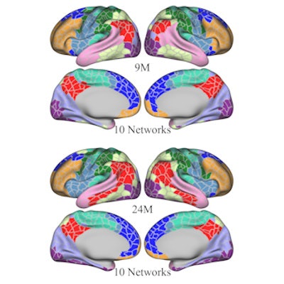
To address the problem, Wang's group created both age-related and age-independent cortical parcellation maps using more than 800 "parcels" (that is, categories of cortical gray matter in different locations in the brain) based on "aligned and averaged local gradient maps of functional connectivity" across the patient cohort.
 Infant-specific fine-grained functional parcellation maps for infants aged from 3 months to 2 years. Images courtesy of Gang Li, PhD. Licensed under CC BY 4.0.
The project included 1,064 resting-state functional MRI scans from 197 infants between the ages of birth to two years; data were collected as part of the University of North Carolina, Chapel Hill/University of Minnesota, Minneapolis Baby Connectome Project Consortium.
The team was successful in creating maps that it hopes will contribute to neuroimaging research -- as they reveal "complex multipeak functions in several aspects, including the across-age variation of local functional gradients, network organization, and local efficiency," it noted.
The study offers perspective that hasn't been part of the literature until now, according to the investigators.
"[Our study provides] the first comprehensive visualizations of infant brain functional developmental maps on the cortex, which will serve as valuable references for future early brain developmental studies," they concluded.
Wang and colleagues have made their infant cortical functional parcellation maps publicly available here, and the complete study can be found here.