Swiss biopharmaceutical company AC Immune highlighted a study showing the potential of its alpha-synuclein (a-syn) PET tracer for diagnosing neurodegenerative disease.
In a first-in-human study published October 27 in Nature Communications, researchers led by Oskar Hansson MD, PhD, of Lund University, found that F-18 ACI-12589 helped visualize specific and reproducible retention pattern in patients with multiple system atrophy (MSA) compared with healthy control subjects.
The finding indicates that the tracer could enable earlier, more accurate diagnosis of MSA and potentially, more precise monitoring of disease progression, AC Immune said.
“Accurately detecting a-syn in the living brains of patients is paramount, particularly in view of the challenges of diagnosing the serious diseases related to pathological a-synuclein aggregates,” noted Marie Kosco-Vilbois, MD, chief scientific officer of AC Immune, in a news release.
A-syn is an abundant, highly soluble unfolded protein that accumulates in the brain most prominently in Parkinson’s disease, dementia with Lewy bodies, and MSA. Unlike in Alzheimer’s disease, for instance, where PET tracers have been developed to visualize beta-amyloid and tau protein accumulations, there is no reliable imaging biomarker available for a-syn.
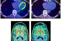
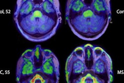
In this study, Hansson and colleagues performed F-18 ACI-12589 PET imaging in 23 participants with a-synuclein related disorders, 11 with other neurodegenerative disorders, and eight controls. The analysis revealed that F-18 ACI-12589 demonstrated clear binding in the cerebellar white matter and middle cerebellar peduncles of MSA patients, regions known to be highly affected by a-synuclein pathology.
 (a) Transversal images at the level of the middle cerebellar peduncles in a control participant, and patients with dementia with Lewy bodies (DLB), multiple system atrophy-cerebellar subtype (MSA-C), and Parkinson’s disease (PD). (b) Transversal images at the level of the basal ganglia in a control participant, and patients with DLB, MSA-parkinsonian type. SUVR images for (a, b) have been created using the occipital cortex as a reference region. Image courtesy of Nature Communications.
“Our results indicate that a-synuclein pathology in MSA can be identified using F-18 ACI-12589 PET imaging, potentially improving the diagnostic work-up of MSA,” the researchers wrote.
The results represent a major step for AC Immune in its further goal to develop precision medicine for neurodegenerative diseases, the company added.
The full article is available here.