An experimental PET radiotracer shows promise for detecting metastatic cancer lesions in patients with neuroendocrine tumors, according to a study published July 20 in the Journal of Nuclear Medicine.
A group led by Zefang Lin, MD, of Fujian Medical University in Fuzhou, China, compared two gallium-68 (Ga-68)-labeled PET radiotracers -- a U.S.-approved agonist type and an experimental antagonist type -- that visualize somatostatin receptor (SSTR) activity in neuroendocrine tumors. They suggest the new tracer has an edge.
"The new radiopharmaceutical Ga-68 NODAGA-JR11 is more valuable than SSTR agonists in patients with NETs, especially in patients with liver-dominant metastases," the group wrote.
SSTRs are overexpressed in certain cancer cells and are valuable targets for PET imaging and for diagnosing neuroendocrine tumors. Ga-68 DOTATATE is an agonist radiotracer approved for this use in the U.S. in 2016.
Agonist and antagonist tracers differ primarily in what happens when they bind to their targets, the researchers explained. Agonist tracers activate SSTRs and are internalized by the target cells. Thus, they provide detailed cellular functional information related to tumors. Meanwhile, antagonist tracers remain predominantly on cell surfaces, and are thought to provide more detail on quantities and the distribution of tumor cells.
According to the researchers, preclinical studies indicate that SSTR-expressing tumors demonstrate higher uptake of antagonist PET tracers than agonists. In this study, they analyzed initial results from an ongoing clinical trial pitting the two types of tracers head-to-head in patients with metastatic, well-differentiated NETs.
The first 48 patients enrolled in the trial (out of 100 planned) between August 2020 and November 2021 received Ga-68 DOTATATE on the first day and experimental Ga-68 NODAGA-JR11 on the second day. Whole-body PET/CT scans were performed between 40 and 60 minutes after injection, with the researchers then comparing normal-organ uptake, lesion numbers, lesion uptake, and sensitivity between the tracers.
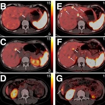
 Ga-68 DOTATATE and Ga-68 NODAGA-JR11 PET/CT images are shown of a postoperative gastric neuroendocrine tumor patient with multiple liver metastases. (A) Ga-68 DOTATATE maximum-intensity projection shows several liver lesions with high uptake. (B) Transaxial fusion image appears to show abnormal moderate-uptake lesion (arrow) in high liver background uptake. (C and D) One transaxial fusion image (C) shows liver metastasis (arrow), whereas no liver lesion was found in another (D). (H) Ga-68 NODAGA-JR11 maximum-intensity projection shows many more liver lesions than Ga-68 DOTATATE. (E-G) Transaxial fusion images at same three levels as in B-D similarly found liver lesions that matched those on Ga-68 DOTATATE (solid arrows). Ga-68 NODAGA-JR11 PET/CT shows lower liver background uptake and additional liver metastases (dashed arrows) at sites that were negative on Ga-68 DOTATATE PET/CT. Image courtesy of the Journal of Nuclear Medicine.
Overall, Ga-68 NODAGA-JR11 demonstrated lower background uptake in normal organs and detected significantly more liver lesions than Ga-68 DOTATATE (673 vs. 584, p = 0.002), the researchers found.
In addition, the group performed a subset analysis in 15 patients in whom 180 lesions had been detected on conventional imaging (MRI or CT) within one year of the study. Of these, 165 were positive on Ga-68 NODAGA-JR11 and 139 were positive on Ga-68 DOTATATE. This calculated to a sensitivity of 91.7% for Ga-68 NODAGA-JR11 and 77.2% for Ga-68 DOTATATE, according to the findings.
"Ga-68 NODAGA-JR11 shows better sensitivity and a higher target-to-background ratio than Ga-68 DOTATATE," the group wrote.
For primary tumors, bone metastases, and lymph node metastases, however, the two tracers were comparable on both patient-based and lesion-based comparisons, the researchers noted.
Ultimately, SSTR-PET imaging plays a critical role in the management of NETs, including staging, restaging, prognosis, therapy decision-making, and response monitoring, the authors noted. This study adds to evidence that has facilitated a shift in the use of SSTR agonist tracers for these purposes to antagonist tracers, despite their lack of internalization into tumor cells, they wrote.
"The results favor Ga-68 NODAGA-JR11 because of a higher detection ability and better image contrast for liver metastases," Lin and colleagues concluded.
A link to the full study can be found here.