ANAHEIM, CA - Researchers have developed a tau PET radioligand that can accurately differentiate in vivo between progressive supranuclear palsy (PSP) and similar brain disorders, according to a study presented Saturday at the Society of Nuclear Medicine and Molecular Imaging (SNMMI) annual meeting.
The researchers from Germany found that PET images showed significantly elevated levels of F-18 PI-2620, which binds to tau proteins in brain regions associated with degenerative neurological diseases. If additional trials prove successful, the radioligand could help diagnose progressive supranuclear palsy in living patients, rather than waiting for an autopsy to confirm the condition.
"The finding indicates great potential for this radioligand to detect an excess of PSP pathology in vivo, specifically for tau deposits with this disease, and to establish an early and more reliable diagnosis of PSP in vivo," said presenter Dr. Matthias Brendel, from the department of nuclear medicine at the University of Munich.
PSP's complexity
Progressive supranuclear palsy is a very aggressive neurodegenerative disease characterized by posture instability and falls, blurred vision, slurred speech, and cognitive decline. Identifying the condition can be challenging and complex since its symptoms overlap with other diseases, such as Parkinson's and Alzheimer's diseases and also cardiovascular degeneration.
"That's why it is not so easy to assess PSP, especially at its early stages," Brendel told SNMMI attendees. "We have low initial sensitivity for differentiating PSP from Parkinson's disease and low specificity for pathological entities."
Also, there is no cure for the condition, and a definitive diagnosis can only be accomplished at autopsy. Therefore, a "good biomarker would provide early diagnosis in a presymptomatic stage," he noted.
This study is not the first time a radioligand has shown some ability to bind to tau deposits to distinguish patients with progressive supranuclear palsy from healthy controls. PET images with F-18 THK5117 received SNMMI's Image of the Year award in 2014. The early results from F-18 PI-2620, however, appear even more encouraging in terms of an in vivo PSP diagnosis.
Tau binding
Brendel and colleagues evaluated nine male and eight female patients (mean age, 70 ± 7 years) with suspected cases of progressive supranuclear palsy. The 17 patients underwent F-18 PI-2620 PET scans at four different imaging centers, along with 10 healthy controls and seven disease controls who had been diagnosed with multisystem atrophy, Parkinson's disease, or Alzheimer's disease.
PET images were acquired within 60 minutes after injection of F-18 PI-2620 and then coregistered to an MRI template. The researchers also calculated standardized uptake value ratios (SUVr) of key brain regions in the basal ganglia and dentate nucleus, including the globus pallidus, substantia nigra, and subthalamic nucleus. They used the cerebellum as the reference region.
Additionally, disease severity was measured by the PSP rating scale, which was correlated with PET findings.
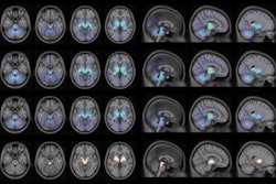
 F-18 PI-2620 binding is easily discernable (red and yellow) in axial and sagittal slices from PSP patients (upper row) and healthy controls (lower row) in an MRI template. Images courtesy of Dr. Mattias Brendel et al and SNMMI.
Time to bind
One noteworthy finding was that the radioligand had "nearly stable binding" of up 20 minutes of scan time, which would enhance the validity of the results, Brendel said.
He and his colleagues also observed significantly greater mean SUVr of F-18 PI-2620 in PSP patients' globus pallidus (1.34 ± 0.16; p = 0.001) and the substantia nigra (1.33 ± 0.14; p = 0.003) compared with healthy controls (1.12 ± 0.09 and 1.15 ± 0.08, respectively).
They found no statistically significant difference in mean SUVr in the globus pallidus (1.11 ± 0.06) and only a slight elevation in the substantia nigra (1.23 ± 0.09) among the disease control subjects compared with the healthy controls. The commonality in these two groups would suggest that F-18 PI-2620 was able to distinguish between patients with PSP and individuals with another form of neurodegeneration.
Interestingly, patients with low PSP disease severity expressed statistically significant elevated levels of F-18 PI-2620 uptake (1.38 ± 0.13) in the globus pallidus compared with the healthy controls (1.12 ± 0.09) (p = 0.001), which again would indicate the radioligand's ability to bind to PSP-related tau deposits.
"The results of this preliminary multicenter evaluation indicate a value of F-18 PI-2620 to diagnose and differentiate suspected PSP patients in vivo," Brendel and colleagues concluded in their SNMMI abstract. They added that the radioligand "may show potential as a biomarker to assess tau pathology in PSP patients and that it may be helpful to establish earlier and more reliable diagnosis of PSP."