Neutron-based molecular imaging could reveal breast cancer at the most basic level, long before anatomical changes are seen, according to a presentation Wednesday at the American Association of Physicists in Medicine (AAPM) conference in Pittsburgh.
Neutron-stimulated emission computed tomography (NSECT) noninvasively maps the concentration of stable or radioactive isotopes in any selected 3D volume of the body, explained Carey Floyd, Ph.D., and colleagues from Duke University in Durham, NC. Floyd is a professor of radiology and biomedical engineering at Duke. His co-authors include Calvin Howell, Ph.D., professor of physics at Duke and deputy director of the Triangle Universities Nuclear Laboratory, where NSECT development is being conducted.
"It is possible that NSECT could play a significant role in (breast cancer) screening. In this application, the whole breast would be illuminated with a very low dose of neutrons," wrote Floyd in an e-mail interview with AuntMinnie.com. "There is evidence from previous research on elemental biodistribution research which suggests that the concentration of trace elements is significantly lower in the whole breast of patients who do not have breast cancer. If NSECT is sensitive to this decrease in trace elements, then it could be used to dramatically reduce the number of women with no disease who report for routine screening mammography without missing cancers."
NSECT imaging by using the inelastic scattering of fast neutrons (1-10 MeV) to differentiate between benign and malignant tissue. The body is illuminated with these neutrons, which as they collide in the body deposit excess energy in the nuclei of atoms and molecules. This excess energy is then released as gamma-ray photons, which are recorded by a detector. The system then reconstructs the locations and energies of the photons, enabling the creation of 3D maps that show the concentration of isotopes throughout the body. The researchers believe that such concentration can occur long before cancer causes the anatomical changes that are detected by modalities like CT and MRI.
 |
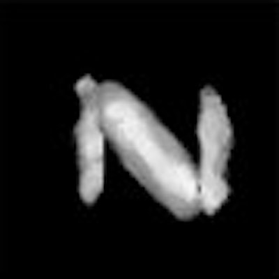
NSECT image (above) and diagram (below) of an iron-copper sample. Striped bars are copper with length of 6 cm, height of 3 cm, and a thickness of 0.7 cm. Solid bars are iron with the same dimensions. The maximum dimension of the sample is 9 cm.
 |
"Spatial projections of the emitted energy spectra allow tomographic image reconstruction of the elemental concentrations," the group wrote in an accompanying abstract. "Malignant regions are identified by artificial neural-network classification of the spectra at each reconstructed image voxel."
In their early experiments, the researchers successfully tested a 7.5-MeV neutron beam on multi-element phantoms containing trace elements of oxygen, carbon, copper, iron, and calcium. They found that images of clinical significance can be achieved with a dose less than that of a standard mammogram.
"This implementation of NSECT would require little dose, could be performed with the woman fully clothed, and could be interpreted very simply from a single (trace element) number.... Depending on the sensitivity and specificity that NSECT is able to provide, it could either reduce the number of screening studies that are performed on women who have low probability of cancer, or could increase the time allocated to women identified with higher risk, or both," Floyd said.
In addition to reduced radiation, NSECT has the edge over other imaging modalities, both molecular and non-molecular. MR spectroscopy (MRS) is limited to a specific set of isotopes, while x-ray fluorescence is best in thin samples or shallow-depth tissues. NSECT can image over the full thickness of the body, the investigators said.
 |
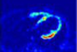
| The image reconstructed from only those photons whose energies were characteristic of copper. Images courtesy of Carey Floyd, Ph.D. |
All of the components required to build an NSECT scanner are currently available. The cost of assembling a prototype scanner would require one-quarter of the capital needed to buy a full CT system, they said. Finally, the square footage required to house an NSECT scanner would be similar to that of an MR unit.
The group cautioned that NSECT is still in the early stages of development: a stand-alone system is expected to be ready for clinical trials within the next five years. A technical drawback of whole-body NSECT, however, is that it is time-consuming. Image interpretation is another issue that may prove controversial. The data generated by an NSECT exam would not require expert interpretation, as the exam result is displayed as a single number, Floyd said. But because many of these patients may still require follow-up imaging with established modalities, mammographers wouldn't necessarily be out of a job, he added.
Floyd said he has high hopes for NSECT, not only for breast cancer screening, but as a diagnostic exam as well. If a suspicious area has already been identified, NSECT could be configured to specifically evaluate that region and potentially determine whether a biopsy is necessary, he said.
By Shalmali PalAuntMinnie.com staff writer
July 28, 2004
Related Reading
Micro MRI tracks breast cancer metastasis, in mice, May 11, 2004
New method provides individualized cancer prognosis, April 28, 2004
15O-water PET shows persistence of blood flow in chemo-resistant breast tumors, November 28, 2003
Copyright © 2004 AuntMinnie.com