Canadian x-ray manufacturer KA Imaging plans to highlight clinical results regarding the use of its Reveal 35C detector in the intensive care unit (ICU) at the American Society of Emergency Radiology (ASER) meeting to be held in Washington, D.C.
Reveal 35C is KA Imaging's single-exposure, dual-energy, flat-panel x-ray detector for use in fixed, mobile, and portable settings, it said.
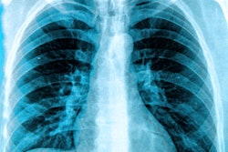
In a study conducted at Grand River Hospital in Kitchener, Ontario, Canada, a team led by Vikram Venkatesh, MD, a diagnostic radiologist in Boston, reported that the detector provides dual-energy images directly at bedside, fixing the problem of transporting ICU patients to imaging when they have multiple lines, tubes, and implantable devices. The group found that study participants reported better image quality, no added reading time, and a reduction in time to treatment/intervention.
"In certain cases, especially when CT is not an option, dual-energy x-ray can permit improved visualization of soft tissues by subtracting unwanted structural noise […] and simultaneously reducing unnecessary radiation exposure," Venkatesh and colleagues wrote. "Medical lines and tubes can be better visualized using the bone image that dual-energy x-ray detectors also produce."
![Representative example of a 16-year-old male patient with underlying X-linked adrenoleukodystrophy. (A, B) Paired anteroposterior (AP) chest radiograph and dual-energy x-ray absorptiometry (DXA) report shows lumbar spine (L1 through L4) areal bone mineral density (BMD). The DXA report was reformatted for anonymization and improved readability. The patient had low BMD (Z score ≤ −2.0). (C) Model (chest radiography [CXR]–BMD) output shows the predicted raw BMD and Z score in comparison with the DXA reference standard, together with interpretability analyses using Shapley additive explanations (SHAP) and gradient-weighted class activation maps. The patient was classified as having low BMD, consistent with the reference standard. AM = age-matched, DEXA = dual-energy x-ray absorptiometry, RM2 = room 2, SNUH = Seoul National University Hospital, YA = young adult.](https://img.auntminnie.com/mindful/smg/workspaces/default/uploads/2026/04/ai-children-bone-density.0snnf2EJjr.jpg?auto=format%2Ccompress&fit=crop&h=100&q=70&w=100)


![Representative example of a 16-year-old male patient with underlying X-linked adrenoleukodystrophy. (A, B) Paired anteroposterior (AP) chest radiograph and dual-energy x-ray absorptiometry (DXA) report shows lumbar spine (L1 through L4) areal bone mineral density (BMD). The DXA report was reformatted for anonymization and improved readability. The patient had low BMD (Z score ≤ −2.0). (C) Model (chest radiography [CXR]–BMD) output shows the predicted raw BMD and Z score in comparison with the DXA reference standard, together with interpretability analyses using Shapley additive explanations (SHAP) and gradient-weighted class activation maps. The patient was classified as having low BMD, consistent with the reference standard. AM = age-matched, DEXA = dual-energy x-ray absorptiometry, RM2 = room 2, SNUH = Seoul National University Hospital, YA = young adult.](https://img.auntminnie.com/mindful/smg/workspaces/default/uploads/2026/04/ai-children-bone-density.0snnf2EJjr.jpg?auto=format%2Ccompress&fit=crop&h=112&q=70&w=112)


