Plain hip x-rays may help screen patients for osteoporosis before they undergo total hip arthroplasty (THA), according to a study published November 14 in Scientific Reports.
The finding is from a study conducted by a group at the University Hospital Jena in Eisenberg, Germany, that explored whether hip x-rays could be reliable indicators for bone mineral density (BMD) in male and female patients prior to THA procedures.
“An easily accessible screening tool for osteopenia or osteoporosis using plain hip radiographs is of great importance for the orthopedic hip surgeons to avoid potential surgical risks while performing total hip arthroplasty due to osteopenia and osteoporosis,” noted lead author Sebastian Rohe, MD, and colleagues.
Studies have shown that dual-energy x-ray absorptiometry (DEXA) is underused in patients prior to THA, despite being the gold standard for assessing osteoporosis, the authors noted. However, several studies have suggested that indices on plain hip x-rays may be reliable screening tools in female or Asian ethnicities.
In this study, the group expanded on this research by investigating the correlation between indices on standardized preoperative x-rays of the hip and the proximal femoral neck T-score, measured by DEXA, in a Caucasian population with a high number of male patients.
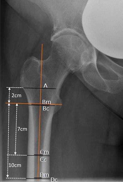
 An example of measurement on plain anterior-posterior x-rays used in the study: canal-bone-ratio CBR-7 cm (Cm/Cc) and CBR-10 cm (Dm/Dc), canal-calcar ratio CCR (Dm/Bm); canal flare index CFI (A/Dm). Image courtesy of Scientific Reports.
The group retrospectively analyzed results from 216 elderly male and female Caucasian patients treated with unilateral THA for primary osteoarthritis and who underwent preoperative DEXA scans and standard x-rays of the hip within one year. They correlated femoral neck DEXA T-scores with four indices on hip x-rays: the canal flare index (CFI), canal calcar ratio (CCR), and canal bone ratio (CBR) 7 cm and 10 cm below the lesser trochanter.
According to the findings, CBR-7 and CBR-10 were highly correlated (p < 0.001) with femoral neck T-score in males and females. In addition, based on an area under the curve (AUC) diagnostic analysis, CBR-7 and CBR-10 also showed good accuracy as metrics for osteoporosis in males (CBR-7: AUC = 0.75; CBR-10: AUC = 0.63) and females (CBR-7: AUC = 0.87; CBR-10: AUC = 0.9).
“Indices such as the CBR 7 or 10 cm below the lesser trochanter on plain hip radiographs are a good screening tool for osteopenia and osteoporosis on plain hip radiographs and can be used to initiate further diagnostics like the gold standard DEXA,” the group wrote.
Ultimately, osteoporosis has become a growing problem in recent years as the number of elderly patients has increased, the authors noted. Furthermore, reflecting the demand for an active lifestyle in older patients, the number of THAs implanted is also increasing, they wrote.
“Looking ahead to the increasing integration of artificial intelligence in the interpretation of plain radiographs, including hip radiographs especially for planning THA, these indices could be used to assess osteopenia or osteoporosis preoperatively and optimize surgical preparation prior to THA,” Rohe and colleagues concluded.
The full article is available here.


