
GE Healthcare has introduced Definium 656 HD, a new generation of its Definium premium fixed x-ray system that features a variety of artificial intelligence (AI)-based functionality.
Utilizing 3D camera technology, GE's Intelligent Workflow Suite is designed to yield more consistent images and avoid the need for retakes, according to the vendor. Technologists can also use a 12-inch touchscreen and GE's AutoRad software to employ automated in-room workflows, as well as manual workflow control, according to the vendor.
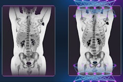
What's more, the overhead tube suspension system on Definium 656 HD supports five-axis motorization and autopositioning, enabling patients to automatically be positioned at any location in the room.
Definium 656 HD. Image courtesy of GE Healthcare.In addition, Definium 656 HD includes GE's 100-micron FlashPad HD detectors and version 2.2 of GE's Helix AI-based image processing software, which delivers anatomy-specific image enhancement regardless of variations in dose, patient positioning, field of view, and the presence of metal implants, the company said.
Other Definium 656 HD features include Auto Image Paste and AutoSpine for stitching of long images, and GE's VolumeRad digital tomosynthesis protocol.
![Representative example of a 16-year-old male patient with underlying X-linked adrenoleukodystrophy. (A, B) Paired anteroposterior (AP) chest radiograph and dual-energy x-ray absorptiometry (DXA) report shows lumbar spine (L1 through L4) areal bone mineral density (BMD). The DXA report was reformatted for anonymization and improved readability. The patient had low BMD (Z score ≤ −2.0). (C) Model (chest radiography [CXR]–BMD) output shows the predicted raw BMD and Z score in comparison with the DXA reference standard, together with interpretability analyses using Shapley additive explanations (SHAP) and gradient-weighted class activation maps. The patient was classified as having low BMD, consistent with the reference standard. AM = age-matched, DEXA = dual-energy x-ray absorptiometry, RM2 = room 2, SNUH = Seoul National University Hospital, YA = young adult.](https://img.auntminnie.com/mindful/smg/workspaces/default/uploads/2026/04/ai-children-bone-density.0snnf2EJjr.jpg?auto=format%2Ccompress&fit=crop&h=100&q=70&w=100)


![Representative example of a 16-year-old male patient with underlying X-linked adrenoleukodystrophy. (A, B) Paired anteroposterior (AP) chest radiograph and dual-energy x-ray absorptiometry (DXA) report shows lumbar spine (L1 through L4) areal bone mineral density (BMD). The DXA report was reformatted for anonymization and improved readability. The patient had low BMD (Z score ≤ −2.0). (C) Model (chest radiography [CXR]–BMD) output shows the predicted raw BMD and Z score in comparison with the DXA reference standard, together with interpretability analyses using Shapley additive explanations (SHAP) and gradient-weighted class activation maps. The patient was classified as having low BMD, consistent with the reference standard. AM = age-matched, DEXA = dual-energy x-ray absorptiometry, RM2 = room 2, SNUH = Seoul National University Hospital, YA = young adult.](https://img.auntminnie.com/mindful/smg/workspaces/default/uploads/2026/04/ai-children-bone-density.0snnf2EJjr.jpg?auto=format%2Ccompress&fit=crop&h=112&q=70&w=112)


