
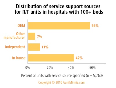
Over half (56%) of the radiography/fluoroscopy (R/F) units installed in U.S. hospitals with 100+ beds are serviced by the manufacturer/OEM, 42% by in-house service, 11% by independent service organizations, and 7% by other manufacturers.

Based on responses to IMV’s 2009/10 Radiographic Fluoroscopy Market Summary Report of U.S. Hospitals with 100+ Beds.
Note: The summation of the percentages by site type exceeds 100% because some respondents specified multiple service support types for their R/F units.
AuntMinnie's IMV MarketStat is provided to AuntMinnie.com by IMV Medical Information Division, Inc. of Des Plaines, IL.

Click here to buy complete IMV Market Reports
MarketStat ArchivesHospital PACS plans for the future
Administration of echo labs
Nonemergency angiography waiting time
Cath lab sites with fixed cath labs
Hard-copy film use at sites with digital mammography units
Nuclear medicine -- Average waiting time
Radiation oncology -- Types of images used in radiation therapy treatment plans
Vascular MRI -- Percent of sites performing and percent of MRI procedures
Single versus multislice detectors in CT installed base
Budgets for nuclear medicine radiopharmaceuticals
Copyright © 2010 AuntMinnie.com
![Representative example of a 16-year-old male patient with underlying X-linked adrenoleukodystrophy. (A, B) Paired anteroposterior (AP) chest radiograph and dual-energy x-ray absorptiometry (DXA) report shows lumbar spine (L1 through L4) areal bone mineral density (BMD). The DXA report was reformatted for anonymization and improved readability. The patient had low BMD (Z score ≤ −2.0). (C) Model (chest radiography [CXR]–BMD) output shows the predicted raw BMD and Z score in comparison with the DXA reference standard, together with interpretability analyses using Shapley additive explanations (SHAP) and gradient-weighted class activation maps. The patient was classified as having low BMD, consistent with the reference standard. AM = age-matched, DEXA = dual-energy x-ray absorptiometry, RM2 = room 2, SNUH = Seoul National University Hospital, YA = young adult.](https://img.auntminnie.com/mindful/smg/workspaces/default/uploads/2026/04/ai-children-bone-density.0snnf2EJjr.jpg?auto=format%2Ccompress&fit=crop&h=100&q=70&w=100)


![Representative example of a 16-year-old male patient with underlying X-linked adrenoleukodystrophy. (A, B) Paired anteroposterior (AP) chest radiograph and dual-energy x-ray absorptiometry (DXA) report shows lumbar spine (L1 through L4) areal bone mineral density (BMD). The DXA report was reformatted for anonymization and improved readability. The patient had low BMD (Z score ≤ −2.0). (C) Model (chest radiography [CXR]–BMD) output shows the predicted raw BMD and Z score in comparison with the DXA reference standard, together with interpretability analyses using Shapley additive explanations (SHAP) and gradient-weighted class activation maps. The patient was classified as having low BMD, consistent with the reference standard. AM = age-matched, DEXA = dual-energy x-ray absorptiometry, RM2 = room 2, SNUH = Seoul National University Hospital, YA = young adult.](https://img.auntminnie.com/mindful/smg/workspaces/default/uploads/2026/04/ai-children-bone-density.0snnf2EJjr.jpg?auto=format%2Ccompress&fit=crop&h=112&q=70&w=112)


