Organizing pneumonia can hide underlying lung cancers in up to 10% of cases, and repeat imaging in the form of PET/CT -- and additional biopsy -- should be considered in patients with high clinical suspicion of malignancy, researchers have reported.
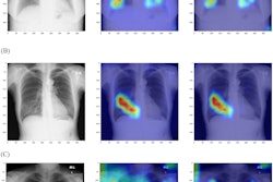
Suspicion of malignancy may be prompted by avid uptake of the radiopharmaceutical FDG on PET/CT imaging, according to study results delivered on November 27 at the RSNA meeting. But it can be tricky to catch potential cancers once organizing pneumonia has been diagnosed, said presenter Charissa Kim, MD, PhD, of Beth Israel Deaconess Medical Center in New York City.
"Accurate diagnosis of organizing pneumonia is confounded by the co-existence of [the condition] with lung malignancies," Kim said.
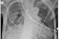
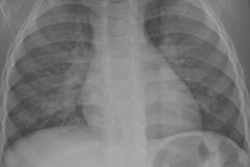
Organizing pneumonia is a form of interstitial lung disease with broad radiologic presentation, she noted, but its features – peripheral consolidation, ground-glass opacities, perilobular opacities, reversed halo sign, and nodules or masses – are nonspecific. She and colleagues evaluated the clinical and imaging features of organizing pneumonia CT-guided biopsy diagnoses that were later shown to be cancer via a study that included 1,314 patients. The patients underwent CT-guided lung biopsy for suspected lung cancer or metastatic disease between February 2014 and April 2022; a pulmonary pathologist confirmed the presence or absence of organizing pneumonia in all samples; the researchers also reviewed chest CTs and/or PET/CTs taken prior to biopsy.
Of the total patient cohort, 98 (7.5%) showed organizing pneumonia on biopsy, with a median lesion size of 2.5 cm. Of these 98 individuals, 10 (10%) of those with this biopsy result underwent another biopsy (at median, 51 days) because of suspicion of malignancy. Pulmonary metastases were found in five of the 10 (50%) and primary lung cancers in the other five (50%).
Kim discussed a case that demonstrated how additional biopsy proved necessary. A 60-year-old woman with hypertension, high cholesterol, gastroesophageal reflux disease, and previous thyroid cancer underwent a PET/CT exam, which showed avid FDG uptake in a lesion in her right middle lobe; this lesion was biopsied and its pathology was interpreted as interstitial pneumonia. However, a six-month follow-up CT exam showed interval growth of the lesion. When it was resected it was determined to be malignant.
"The initial biopsied portion likely represented organizing pneumonia adjacent to malignancy," Kim said.
The bottom line? As both organizing pneumonia and malignancy can be FDG-avid, "repeat biopsy should be considered in patients with high clinical suspicion of malignancy despite an initial pathology diagnosis of organizing pneumonia," she concluded.