
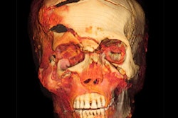
When an international team of researchers acquired micro-CT scans of what they believed was a mummy of an Egyptian hawk, they discovered instead that it was a human fetus.
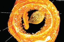
Micro-CT scan of the male fetus. Image courtesy of Maidstone Museum and Nikon Metrology.The researchers, led by bioarchaeologist and mummy expert Andrew Nelson, PhD, of Western University in Canada, examined high-resolution micro-CT scans of the mummy.
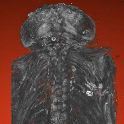
They found that it was a male fetus with several congenital health abnormalities, including anencephaly, a rare condition in which the brain and skull do not develop properly.
The fetus' previous label as a mummified hawk from the Ptolemaic Period has been changed to reflect this discovery at its home in the Maidstone Museum in the U.K.


![Images show the pectoralis muscles of a healthy male individual who never smoked (age, 66 years; height, 178 cm; body mass index [BMI, calculated as weight in kilograms divided by height in meters squared], 28.4; number of cigarette pack-years, 0; forced expiratory volume in 1 second [FEV1], 97.6% predicted; FEV1: forced vital capacity [FVC] ratio, 0.71; pectoralis muscle area [PMA], 59.4 cm2; pectoralis muscle volume [PMV], 764 cm3) and a male individual with a smoking history and chronic obstructive pulmonary disorder (COPD) (age, 66 years; height, 178 cm; BMI, 27.5; number of cigarette pack-years, 43.2, FEV1, 48% predicted; FEV1:FVC, 0.56; PMA, 35 cm2; PMV, 480.8 cm3) from the Canadian Cohort Obstructive Lung Disease (i.e., CanCOLD) study. The CT image is shown in the axial plane. The PMV is automatically extracted using the developed deep learning model and overlayed onto the lungs for visual clarity.](https://img.auntminnie.com/mindful/smg/workspaces/default/uploads/2026/03/genkin.25LqljVF0y.jpg?auto=format%2Ccompress&crop=focalpoint&fit=crop&h=112&q=70&w=112)


