Tuesday, November 29 | 7:15 a.m.-8:15 a.m. | SPSH30 | Room E352
This Tuesday Hot Topic Session will offer a comprehensive review of the technical and clinical aspects of various types of spectral CT imaging, including established techniques such as dual-source and rapid kVp switching, and emerging techniques such as split-beam CT, spectral detector CT, and photon counting.The session will be co-moderated by Dr. Suhny Abbara from the University of Texas Southwest Medical Center and Dr. David Bluemke, PhD, from the U.S. National Institutes of Health.
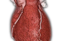
Multispectral CT imaging is increasingly available for a wide range of cardiovascular imaging applications, where it can be used to increase the conspicuity of atherosclerotic plaque and improve the visualization of calcifications.
"Improved energy optimization results in lower radiation dose while maintaining high iodine enhancement levels," Bluemke wrote in an email to AuntMinnie.com. "Overall, contrast-to-noise ratios can be improved for better depiction of myocardial infarction."
Other advantages of multispectral CT include improved material classification for coronary artery plaque and myocardial scar, according to Bluemke. Photon-counting CT is a type of multispectral CT that further offers the potential for improved spatial resolution along with accurate material classification.
Bluemke will lead a talk on current applications in spectral cardiovascular CT, while Abbara will discuss opportunities in spectral detector cardiovascular CT.