A cloud-based platform described as a "virtual workstation" for radiology education was unveiled by Massachusetts General Hospital (MGH) at last week's Society of Cardiovascular Computed Tomography (SCCT) conference in Las Vegas.
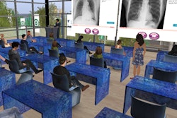
Called RadIQ, the Web-based technology is designed to offer a more immersive educational environment than static images and encyclopedic text, according to its developers, Dr. Garry Choy and Dr. Udo Hoffmann. RadIQ uses a simulated training environment that is similar to a full clinical workstation. The program currently includes 250 cases organized in 10 courses, and it is available at www.RadIQ.org.
One RadIQ program featured at SCCT 2015 was a cardiac CT curriculum directed by Dr. Christopher Maroules. The module is equipped with 50 full cardiac CT datasets and an image viewer that shows users how to manipulate cardiac images as if they were on a 3D workstation.
RadIQ's cardiac CT curriculum is being offered for free to all residents and fellows who become members of SCCT. This gives them the opportunity to finish their level 1 competency in cardiac CT before finishing their training programs, regardless of how much cardiac CT is being performed at their facilities, according to Maroules.
Calgary Scientific provides the viewer embedded in the platform, enabling users to view full DICOM datasets via its resolutionMD viewer.


![Images show the pectoralis muscles of a healthy male individual who never smoked (age, 66 years; height, 178 cm; body mass index [BMI, calculated as weight in kilograms divided by height in meters squared], 28.4; number of cigarette pack-years, 0; forced expiratory volume in 1 second [FEV1], 97.6% predicted; FEV1: forced vital capacity [FVC] ratio, 0.71; pectoralis muscle area [PMA], 59.4 cm2; pectoralis muscle volume [PMV], 764 cm3) and a male individual with a smoking history and chronic obstructive pulmonary disorder (COPD) (age, 66 years; height, 178 cm; BMI, 27.5; number of cigarette pack-years, 43.2, FEV1, 48% predicted; FEV1:FVC, 0.56; PMA, 35 cm2; PMV, 480.8 cm3) from the Canadian Cohort Obstructive Lung Disease (i.e., CanCOLD) study. The CT image is shown in the axial plane. The PMV is automatically extracted using the developed deep learning model and overlayed onto the lungs for visual clarity.](https://img.auntminnie.com/mindful/smg/workspaces/default/uploads/2026/03/genkin.25LqljVF0y.jpg?auto=format%2Ccompress&crop=focalpoint&fit=crop&h=100&q=70&w=100)


![Images show the pectoralis muscles of a healthy male individual who never smoked (age, 66 years; height, 178 cm; body mass index [BMI, calculated as weight in kilograms divided by height in meters squared], 28.4; number of cigarette pack-years, 0; forced expiratory volume in 1 second [FEV1], 97.6% predicted; FEV1: forced vital capacity [FVC] ratio, 0.71; pectoralis muscle area [PMA], 59.4 cm2; pectoralis muscle volume [PMV], 764 cm3) and a male individual with a smoking history and chronic obstructive pulmonary disorder (COPD) (age, 66 years; height, 178 cm; BMI, 27.5; number of cigarette pack-years, 43.2, FEV1, 48% predicted; FEV1:FVC, 0.56; PMA, 35 cm2; PMV, 480.8 cm3) from the Canadian Cohort Obstructive Lung Disease (i.e., CanCOLD) study. The CT image is shown in the axial plane. The PMV is automatically extracted using the developed deep learning model and overlayed onto the lungs for visual clarity.](https://img.auntminnie.com/mindful/smg/workspaces/default/uploads/2026/03/genkin.25LqljVF0y.jpg?auto=format%2Ccompress&crop=focalpoint&fit=crop&h=112&q=70&w=112)


