The International Lymphoma Radiation Oncology Group (ILROG) has issued a guideline on the use of 3D CT-based radiation therapy planning and volumetric image guidance to treat pediatric Hodgkin's lymphoma.
The standards are also designed to reduce radiation dose to normal tissue, decreasing the risk of late side effects. The guideline will be published in the March-April issue of Practical Radiation Oncology, the clinical practice journal of the American Society for Radiation Oncology (ASTRO).
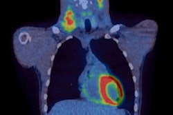
The authors describe methods for identifying target volumes for radiation therapy, as well as how to define radiation target volumes and limit dose to normal organs at risk. The guideline stresses that accurate assessment of the extent and location of disease requires both contrast-enhanced CT and FDG-PET.
The guideline could reduce the radiation therapy breast dose by about 80% and heart dose by about 65% for an adolescent girl with Hodgkin's lymphoma, said lead author Dr. David Hodgson, an associate professor in the department of radiation oncology at the University of Toronto, in a statement from ASTRO.
More personalized treatment planning tailored to an individual's disease will optimize risk-benefit considerations for patients and reduce the likelihood of late effects from radiation therapy, he added.


![Images show the pectoralis muscles of a healthy male individual who never smoked (age, 66 years; height, 178 cm; body mass index [BMI, calculated as weight in kilograms divided by height in meters squared], 28.4; number of cigarette pack-years, 0; forced expiratory volume in 1 second [FEV1], 97.6% predicted; FEV1: forced vital capacity [FVC] ratio, 0.71; pectoralis muscle area [PMA], 59.4 cm2; pectoralis muscle volume [PMV], 764 cm3) and a male individual with a smoking history and chronic obstructive pulmonary disorder (COPD) (age, 66 years; height, 178 cm; BMI, 27.5; number of cigarette pack-years, 43.2, FEV1, 48% predicted; FEV1:FVC, 0.56; PMA, 35 cm2; PMV, 480.8 cm3) from the Canadian Cohort Obstructive Lung Disease (i.e., CanCOLD) study. The CT image is shown in the axial plane. The PMV is automatically extracted using the developed deep learning model and overlayed onto the lungs for visual clarity.](https://img.auntminnie.com/mindful/smg/workspaces/default/uploads/2026/03/genkin.25LqljVF0y.jpg?auto=format%2Ccompress&crop=focalpoint&fit=crop&h=100&q=70&w=100)


![Images show the pectoralis muscles of a healthy male individual who never smoked (age, 66 years; height, 178 cm; body mass index [BMI, calculated as weight in kilograms divided by height in meters squared], 28.4; number of cigarette pack-years, 0; forced expiratory volume in 1 second [FEV1], 97.6% predicted; FEV1: forced vital capacity [FVC] ratio, 0.71; pectoralis muscle area [PMA], 59.4 cm2; pectoralis muscle volume [PMV], 764 cm3) and a male individual with a smoking history and chronic obstructive pulmonary disorder (COPD) (age, 66 years; height, 178 cm; BMI, 27.5; number of cigarette pack-years, 43.2, FEV1, 48% predicted; FEV1:FVC, 0.56; PMA, 35 cm2; PMV, 480.8 cm3) from the Canadian Cohort Obstructive Lung Disease (i.e., CanCOLD) study. The CT image is shown in the axial plane. The PMV is automatically extracted using the developed deep learning model and overlayed onto the lungs for visual clarity.](https://img.auntminnie.com/mindful/smg/workspaces/default/uploads/2026/03/genkin.25LqljVF0y.jpg?auto=format%2Ccompress&crop=focalpoint&fit=crop&h=112&q=70&w=112)


