Monday, November 26 | 11:20 a.m.-11:30 a.m. | SSC14-06 | Room S403A
The clinical applications for digital tomosynthesis have been growing steadily, and Japanese researchers believe that sinus imaging should be added to the list.Abnormal opacity is a telltale sign of paranasal sinusitis, but conventional radiography can be confounded by a number of factors, such as overlying soft tissue, variation in sinus depth, and insufficient image quality. Many institutions routinely use thin-slice MDCT due to its high image quality, but radiation dose -- especially to the lens of the eye -- can be an important issue, according to Dr. Haruhiko Machida of Tokyo Women's Medical University.
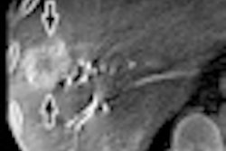
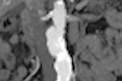
To test the suitability of digital tomo for this application, Machida's team performed sinus MDCT and coronal tomosynthesis in 31 patients (16 men, 15 women; average age, 56.4 years) with suspected acute or chronic sinusitis. Images were reviewed by two readers who determined the presence of opacification, and results were compared between modalities.
Results were assessed for each of the maxillary, ethmoid, frontal, and sphenoid sinuses. Tomosynthesis allowed good detection of opacification, especially in the maxillary, frontal, and sphenoid sinuses, even by readers without many years of experience in interpreting the images, Machida said. In addition, tomosynthesis administered about 1/300th the radiation dose of sinus MDCT, according to the group.