Toshiba Medical Systems Europe has introduced its adaptive iterative dose reduction (AIDR) technology, designed to reduce radiation dose by as much as 75% compared to conventional scanners.
The technology is available with Toshiba's Aquilion One, Aquilion Premium edition, and all 64-detector-row CT scanners.
Images processed with AIDR technology show enhanced image quality with lower noise levels compared to standard protocols using identical dose, Toshiba said. The AIDR technology is adaptive and automatically calculates the optimized number of iterations for any acquisition, according to the company.
Toshiba plans to showcase the technology at the European Society of Cardiology (ESC) Congress in Stockholm at the end of August.
Related Reading
Toshiba taps new MR business unit director, August 12, 2010
Toshiba launches Aegis workstation, June 23, 2010
Toshiba taps ultrasound director, June 17, 2010
Toshiba taps new CEO, June 9, 2010
Toshiba launches V-TRACE for MRA, March 22, 2010
Copyright © 2010 AuntMinnie.com


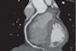
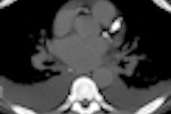


![Axial images from unenhanced calcium score cardiac CT (left) and curved planar reformation images from CT angiography (right) show that higher long-term exposure to air pollution is associated with greater coronary artery calcium and more obstructive coronary artery disease (CAD). Top row: Images in a 68-year-old male patient with higher 10-year mean ambient air pollution exposure (7.9 μg/m3 for particulate matter measuring ≤2.5 μm in diameter [PM2.5] and 17.4 parts per billion [ppb] for NO2) with extensive CAD (coronary artery calcium score [CACS] >1,000 and obstructive CAD [≥70% diameter stenosis]). Bottom row: Images in a 57-year-old female patient with lower 10-year mean ambient air pollution exposure (6.3 μg/m3 for PM2.5 and 4.6 ppb for NO2) with no CAD (CACS = 0 and no obstructive stenosis).](https://img.auntminnie.com/mindful/smg/workspaces/default/uploads/2026/06/hanneman.r6SMLzkezo.png?auto=format%2Ccompress&dpr=2&fit=crop&h=167&q=70&w=250)


