Dear CT Insider,
CT angiography practices in community-based imaging centers are rare birds indeed. But as detector rows multiply like rabbits, and CTA keeps earning its stripes as an alternative to conventional angiography, we can only expect to see more of them popping up on the medical landscape.
How to set up and run such a practice is the million-dollar question, with so few examples to navigate by. One practice that bears a closer look is Atlantic Medical Imaging in Galloway, NJ, which has built a successful business model around its CTA exam.
In the first installment of our two-part series on the community-based CTA practice, radiologist and imaging center proprietor Dr. David Dowe focuses on the patient selection process, designed to accommodate the would-be patient as well as the bottom line.
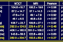
Some patients simply shouldn't be scanned in the outpatient setting, of course. In a talk last month at the International Symposium on Multidetector-Row CT, sponsored by Stanford University in California, Dowe discussed which ones should still be imaged at the hospital, even when the outpatient practice is equipped with a 64-slice scanner.
Once the patient selection hurdle has been cleared, workflow planning becomes a straightforward process, and the practice can offer both high throughput and a high level of service for the patient, according to Dowe.
Find out how Atlantic Medical Imaging does it in this issue's CT Insider Exclusive, made available exclusively to Insider subscribers before it is published on our general site. And be sure to check back for part II, wherein Dowe explains how to build a CTA practice from the ground up, all in your CT Digital Community.