Best Radiology Image
Minnies 2026 winner: Brain PET scans reveal neuron loss in long COVID. Image from Jeffrey Meyer, MD, PhD, of the Centre for Addiction and Mental Health (CAMH) in Toronto, Ontario.
For years, the neuropsychiatric symptoms of long COVID, such as brain fog, apathy, slowed movement, and memory problems, have been elusive to researchers. A PET imaging study from the Centre for Addiction and Mental Health (CAMH) in Toronto produced direct visual evidence of what may be driving them.
The image, which shows roughly 20% lower vesicular monoamine transporter 2 (VMAT2) binding in long COVID patients compared to healthy controls, is the Minnies' Image of the Year.
“This study discovered strong evidence for loss of dopamine-releasing neurons in long COVID,” said senior author Jeffrey Meyer, MD, PhD, a senior scientist with the Brain Health Imaging Centre at CAMH. “PET imaging with VMAT2 binding radiopharmaceuticals would be an optimal way of detecting long COVID with neuropsychiatric symptoms, either for treatment development or in clinical settings as future treatments for long COVID are established.”
 Representative participant from each group shown. C-11 DTBZ non-displaceable binding potential (BPND) in voxels quantitated with simplified reference tissue model 2. Color scale indicates C-11 DTBZ BPND from 0 (black) to 3.5 (red), with reduced C-11 DTBZ BPND observed in the long COVID case compared to both healthy controls. eBioMedicine
The images were acquired on a 3D, high-resolution research PET scanner using a radiotracer called carbon-11 (C-11) dihydrotetrabenazine (DTBZ), which binds to VMAT2, a protein on the nerve endings of dopamine-releasing neurons that serves as a marker of their density. Binding values were derived using the simplified reference tissue model 2, an approach that reduces the influence of blood flow on the measurement and focuses more directly on tracer binding, Meyer explained. VMAT2 binding was compared across three groups: long COVID patients experiencing neuropsychiatric symptoms, healthy controls scanned before the pandemic, and healthy controls who had previously had COVID-19 but recovered fully.
The magnitude of the difference surprised even Meyer. “Prior to the study, I had hypothesized conservatively a difference of 10% to 15%, but being the first study to explore this question, you don’t truly know what you will find until you do the study,” Meyer said.
The pattern of binding loss also varied meaningfully by brain region, and each deficit mapped onto a specific cluster of symptoms. Loss in the ventral striatum correlated with apathy; loss in the dorsal putamen with motor slowing; and loss in the dorsal caudate with decreased verbal memory. The regional specificity of those findings has direct implications for treatment, Meyer added.
“This argues for developing therapeutics to restore or augment function of dopamine-releasing neurons,” he said.
The imaging approach could also take on a diagnostic or stratification role, according to Meyer. PET scanning with VMAT2-binding radiopharmaceuticals could be used to identify long COVID patients with neuropsychiatric involvement in clinical settings, or to enrich clinical trials as therapies targeting this pathway are developed, he noted. The scanner used in the study offers somewhat higher spatial resolution than conventional clinical PET systems, and the SRTM2 modeling approach is a refinement not typically applied in routine clinical practice, and these factors that could inform how the method is eventually adapted for wider use, Meyer suggested.
“There were also correlations between regional loss in ventral striatum, dorsal putamen and dorsal caudate with symptoms of apathy, motor slowing and decreased verbal memory respectively,” he said. “This argues for developing therapeutics to restore or augment function of dopamine-releasing neurons.”
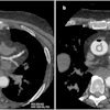
Runner up: Cinematic rendering of penetrating cardiac injury. Image from Elliot Fishman, MD, of Johns Hopkins University.
Scientific Paper of the Year
Minnies 2026 Winner: The effect of AI on the Radiologist Workforce: A Task-Based Analysis. Curtis Langlotz, MD, PhD. medRxiv, December 2025. To learn more about this paper, click here.
In a rarity, the Minnies expert panel has selected a paper published on medRxiv.org as the winner of Scientific Paper of the Year. Perhaps because it’s a deep dive into AI’s impact on radiology by one of the luminaries in the field, Curt Langlotz, MD, PhD, of Stanford University.
The paper projects that AI could reduce radiologist hours worked by as much as 49% over five years, but that sustained growth in imaging volumes will likely absorb much of that effect, with the net result being a shift in what radiologists do rather than a reduction in how many are needed.
The roots of the work go back a decade. In 2016, many experts were predicting that AI would replace radiologists. Langlotz was concerned about how that would affect medical student decisions to choose radiology as a profession. That led to a commentary published in Radiology: Artificial Intelligence, which made a rhetorical argument for why radiologist replacement was unlikely. By late 2024, he decided it was time for something more rigorous.
“I realized that for many radiology tasks, a few studies did provide solid data on those effects,” he told AuntMinnie.com. “So I pushed to make the prediction less rhetorical and more quantitative.”
Economists have long used task-based analysis to study technology’s effect on the workforce, and Langlotz applied that framework to radiology, mapping how radiologists spend their time and then estimating, task by task, where AI tools are likely to make an impact.
The two biggest levers he identified were report drafting and study delegation. Langlotz said he expects report drafting, where AI generates a first draft for the radiologist to review and sign, to arrive first.
“I believe report drafting will drive adoption more rapidly because it saves time,” Langlotz said. “Nobody likes starting with a blank page. And I think report drafting is a relatively safe work process. Academic radiologists routinely review an imaging study and a draft report together to make sure the report is correct before they sign it. The only difference is the draft report is created by a human trainee, not AI.”
Study delegation, such as routing studies that AI flags as almost certainly normal to AI-only interpretation, is a tougher sell, as prospective trial evidence will likely be required before that model sees wide adoption, he said.
“Radiologists are taught that every study must be reviewed by a radiologist to avoid the risk of a missed abnormality,” Langlotz said. “So it goes against our lifelong training to delegate some images to AI-only interpretation.”
He added that AI tools will eventually need to be introduced to radiology residents, though the right moment is an open question.
“Nobody believes trainees should use report drafting tools from day one — just like we don’t give calculators to elementary school children who are learning their multiplication tables,” he said.
Langlotz is currently working with a radiology resident and two computer science graduate students, both trained as physicians, to develop a more defensible version of the paper using formal literature reviews and expert panels. He said his decision to post on medRxiv, however, was deliberate.
“A couple of journals were willing to consider publishing it as a commentary,” he said, “But that usually came with severe limitations on length, figures, and citations that I felt detracted too much from the message.”
Runner up: Radiologist turnover in the United States. Parikh, et al, Journal of the American College of Radiology, June 23, 2026.
Best New Radiology Device
Minnies 2026 winner: Photonova Spectra, GE HealthCare
The introduction of GE HealthCare’s first photon-counting CT scanner was a significant milestone for the company, reflecting years of focused investment in advancing CT technology, according to Chad Rowland, executive director, global CT.
Featuring the firm’s Deep Silicon detector technology, Photonova Spectra combines ultrahigh-definition spatial resolution and spectral imaging capabilities in a single acquisition.
“It reflects our belief that the future of CT is not simply about seeing anatomy in greater detail, but also understanding more about the tissues and structures being imaged,” Rowland told AuntMinnie. “By capturing both spatial and spectral information, Photonova Spectra is designed to give clinicians greater flexibility to investigate complex findings and support more informed clinical assessments. Equally important is making these advanced capabilities practical for everyday clinical use.”
GEHC is highlighting the benefits of Photonova Spectra’s universal one-scan workflow, which captures both spatial and spectral information without requiring clinicians to anticipate every imaging question before an exam begins, according to Rowland.
“The goal is to provide rich information without introducing unnecessary workflow complexity,” he said.
 GE HealthCare's Photonova Spectra photon-counting CT scanner.GE HealthCare
After receiving U.S. Food and Drug Administration (FDA) 510(k) clearance in March, Photonova Spectra has since received the CE Mark in Europe as well as regulatory approval in Japan. Since the scanner’s launch, GEHC has also continued to expand its clinical and research collaborations to explore the technology’s capabilities and potential applications, Rowland said.
“This work brings together leading institutions across North America, Europe and Asia to evaluate image quality, spectral performance, quantitative imaging, clinical utility and workflow considerations,” he said. “These collaborations include research in cardiovascular imaging, tissue characterization, ultra-high definition visualization and imaging protocol optimization. By working closely with clinicians, researchers and medical physicists, GE HealthCare aims to generate additional clinical and technical evidence that can help inform the continued evolution of Photonova Spectra.”
AuntMinnie.com · Minnies 2026 - Best New Radiology Device
Runner-up: uMI Panvivo Mobile PET/CT scanner, United Imaging
Best New Radiology Software
Minnies 2026 winner: AiMIFY, Bracco Imaging/Subtle Medical
This year’s Minnies winner is the first AI software cleared by the U.S. Food and Drug Administration that amplifies brain lesion MR image contrast following administration of a gadolinium-based contrast agent (GBCA). Developed by Bracco in collaboration with Subtle Medical, AiMIFY virtually increases contrast enhancement up to a level that would be expected from a double dose of a Bracco GBCA without needing the extra dose of contrast, according to Bracco.
In a global collaboration that began in September 2023, Bracco and Subtle Medical have been working together to develop AI technology for contrast-enhanced MRI.
“This collaboration leverages the two companies’ expertise in their respective domains: contrast imaging for Bracco and AI-powered image acquisition for Subtle Medical,” a Bracco spokesperson said.
The result is software that can support diagnostic confidence for neurologists in challenging clinical cases, by improving visualization of small lesions, and by improving quantitative image-quality metrics of contrast-enhanced brain images, Bracco said. It averages a 100% increase in contrast enhancement percentage. The firm noted that AiMIFY can also be seamlessly integrated into clinical workflow. Bracco Imaging
AiMIFY received U.S. Food and Drug Administration clearance in 2024 and received the CE Mark in Europe in June 2025. And in November the software was approved for use in Brazil.
“Customer feedback on AiMIFY has been positive, with radiologists reporting that AiMIFY processed images can make enhancing lesions easier and faster to visualize, while reducing image noise and motion artifacts,” a Bracco spokesperson said. “Early pilot experience is now progressing into commercial adoption at U.S. institutions.”
AuntMinnie.com · Minnies 2026 - Best New Radiology Software
Runner-up: Rapid Aortic aortic disease assessment software, RapidAI
Best New Radiology Vendor
Minnies 2026 winner: Shadowfax AI
For Best New Radiology Vendor Shadowfax AI, it’s all about providing radiologists with software that respects their time, according to CEO Reed Branson.
“Imaging informs decisions throughout medicine, but the tools surrounding image interpretation can make radiologists’ work unnecessarily difficult,” Branson said. “Slow interfaces, fragmented applications, repetitive reporting tasks, and workflow interruptions consume time and attention that could otherwise go toward patient care. Shadowfax brings together practicing radiologists and experienced engineers to address those problems.”
Shadowfax AI has built a cloud-based radiology software platform that provides workflow orchestration, image viewing, reporting, mobile dictation, and operational analytics, according to the vendor.
“Practicing radiologists guide product development, with an emphasis on speed, thoughtful design, and the realities of the reading room,” Branson said.
 Shadowfax’s Prism reporting workspace brings patient context, prior-study findings, comparison tools, and report editing into a single interface. Demonstration case with fictional patient information.Image and caption courtesy of Shadowfax.
He said that the vendor approaches the reading workflow as a connected experience, from selecting the next study through reviewing images, creating a report, and understanding the practice’s operations.
“Our goal is software that is fast, intuitive, and enjoyable to use, with AI built into the tasks where it can be useful,” Branson told AuntMinnie. “Throughout the platform, physicians retain clinical control. AI supports report preparation and workflow, while radiologists interpret the images, review the output, and make the final clinical decisions.”
After debuting its Prism AI-augmented reporting application at RSNA 2025, Shadowfax launched its integrated radiology suite in June. In July, the vendor unveiled Shadowfax Mobile, an iOS and Android app that enables radiologists to use their smartphones for dictation and reporting controls. And in August, Shadowfax announced it would provide eligible radiology residents, fellows, and other radiologists in training with free access to Prism and Shadowfax Mobile for the duration of their training.
AuntMinnie.com · Minnies 2026 - Best New Radiology Vendor
Runner-up: Vista AI
Best Educational Mobile App
Minnies 2026 winner: MagnetVision Pro, Emanuel Kanal, MD, (iOS)
As a licensed instrument-rated pilot in addition to being a radiologist, Emanuel “Manny” Kanal, MD, modeled his approach to MR safety on what he learned from the extensive safety-related activities that he observed in the aviation industry.
He designed MagnetVision Pro to serve as an advanced clinical MR safety simulator while also simultaneously implementing his “Kanal Method” of using a custom-developed checklist for risk evaluation of patients in whom there might be implants, devices, and/or foreign bodies prior to clearing them to undergo a requested MR examination.
“I used this high-resolution simulator and checklist software as the backbone of my 26-hour long MR safety courses that I first created back in 2014,” Kanal told AuntMinnie. “To date, more than 10,000 radiologists, technologists, physicists, veterinarians, and various types of physicians and researchers have attended these courses and have used or seen me use this software in these courses that I have given in more than two dozen cities and countries throughout the world.”
 Top of GE 450W 1.5T MR scanner rendered invisible with 6’ tall 70 kg patient positioned in the bore for an MRI examination of the brain. The strength and spatial distribution of the transmitted radiofrequency oscillating magnetic fields of an unloaded body coil for this model MR scanner color coded are superimposed on our “patient" to demonstrate the areas of highest and lowest direct RF-irradiation exposure levels. This helps explain why, when using the body RF coil for RF transmission for a brain MRI examination, the patient can develop a severe proximity at the upper outer aspects of the shoulder and arms where there is intense exposure to direct B1 (RF) irradiation intensities (shown in white) and, therefore, strong induced electrical fields and currents.Image and caption courtesy of Emanuel Kanal, MD.
The app has been significantly expanded in scope and capabilities over the last few years, according to Kanal.
“Instead of being a purely educational tool, the app has now massively grown into a product designed to safely and efficiently run MR imaging centers with special focus on being able to safely yet highly efficiently run remote MR imaging," he said. "‘The most expensive MR scanner is an empty one.’ Unanticipated implants, devices, foreign bodies, etc. in patients showing up for their MR examinations is a leading cause for delays and inefficiencies in clinical MRI today.”
Custom-developed safety algorithms have also been incorporated into MagnetVision Pro.
“This enables it to automatically analyze patient implants, body habitus, and other patient safety information to help improve not only patient safety but simultaneously notably increasing site efficiency for clinical - and especially for remote - MR scanning,” he said.
AuntMinnie.com · Minnies 2026 - Best Educational Mobile App
Runner-up: IMAIOS e-anatomy, IMAIOS (iOS)
Continue reading: Back to Page 1, Page 2