Dear AuntMinnie Member,
The spring radiology conference season was in full swing this week. The International Society for Magnetic Resonance in Medicine (ISMRM) and the International Society for MR Radiographers and Technologists (ISMRT) held their annual meeting and exhibition in Singapore, while the American Roentgen Ray Society (ARRS) hosted their 124th annual meeting in Boston.
Not surprisingly, coverage from those two meetings dominated our list of most highly viewed articles from last week. And a video interview from ISMRM 2024 with Charlene Liew, MD, director of cardiothoracic imaging at Changi General Hospital in Singapore, was in the top spot.
Liew, who had given a plenary talk earlier in the conference, described what she called the four horsemen of the radiology AI apocalypse – the key obstacles to integrating AI into the radiology department – and offered advice on how to address them.
Our third-most popular story included a video interview with ultrahigh-field MRI expert Nicolas Boulant, PhD, of the NeuroSpin research center in France. Click here to learn about his team’s early clinical experience with neuroimaging 20 subjects using 11.7-tesla MRI.
Other highly viewed articles from ISMRM 2024 featured research on how MR spectroscopic imaging can show the effects of HIV treatment on the brain and a discussion on the role of MRI in the emergency department.
Be sure to stop by our ISMRM 2024 RADCast for over 30 articles and video interviews.
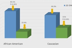
Meanwhile, our second-most popular article this week included coverage of a presentation at ARRS 2024 on the potential complications that can occur during theranostics treatments. Also drawing attention was a story on how digital breast tomosynthesis (DBT) can result in fewer breast cancer screening recalls among African American women.
Researchers have found consistent disparities among different races and sexes for the risk of not matching into diagnostic radiology residency programs, according to another ARRS 2024 presentation. But there are signs of improvement.
See the full list below of our most popular stories of the week:
- ISMRM: Beware the ‘four horsemen of the radiology AI apocalypse’
- ARRS: How to manage the side effects of radiopharmaceuticals
- ISMRM: 11.7-tesla MRI: ‘The Good, the Bad, and the Ugly’
- ARRS: DBT leads to fewer recalls among African American women
- ISMRM: Radiology AI technology is here – and more is coming
- ISMRM: MRI shows effects of HIV treatment on the brain
- ISMRM: Does MRI have a role in the emergency department?
- ARRT awarded $50K in Tennessee RT case
- ISMRM: Advanced MRI techniques shed new light on multiple sclerosis
- ISMRM: Technologist inexperience leads to MRI accidents
- ISMRM: Pinker-Domenig discusses her research, the ‘buzz’ on AI, trauma imaging
- ARRS: Consistent disparities found in radiology residency match
- ISMRM: Is low-field MRI coming of age for lung imaging?
- ACR responds to Congressional query on AI reimbursement

![Examples of ultrasound findings and techniques. (A) Images in a 39-year-old male patient with a mass in the left thigh. The mass is heterogeneous on the B-mode US image (compared with the patient in D) and showed increased microvascularity (superb microvascular imaging [SMI]) and shear-wave elastography (SWE) values. Undifferentiated pleomorphic sarcoma was diagnosed at biopsy (with pleomorphic rhabdomyosarcoma in surgical specimen). (B) Images in an 18-year-old male patient with a mass in the left leg. The mass is hypoechoic on the B-mode image, with no other findings suggestive of malignancy. The lesion is in contact with the cortex of the tibia, which is slightly irregular. CT revealed a doubtful anteromedial tibial erosion. The microvascular study demonstrated high vascularization, suggestive of malignancy. Periosteal Ewing sarcoma was diagnosed with both histologic and immunohistochemical confirmation. (C) Images in a 69-year-old female patient with a lump growing on the outside of the left leg. Multiple SWE examinations were performed (please note the high values obtained in the measurements, whereas the color map highlights the stiffness relative to adjacent tissues). SMI showed areas of increased vascularization to target for sampling. Undifferentiated spindle cell sarcoma was diagnosed at biopsy, with residual leiomyosarcoma in the surgical specimen after neoadjuvant therapy. (D) Images in a 56-year-old female patient with a mass in the right thigh. The mass is heterogeneous at both B-mode ultrasound (similar to patient A) and MRI (coronal T2-weighted spectral attenuated inversion recovery [SPAIR]; T1-weighted pre-contrast and postcontrast imaging), which even shows uptake after the administration of paramagnetic contrast material, which is traditionally suggestive of malignancy. Low values at SMI and elastography are suggestive of benignity. Spindle cell lipoma was diagnosed at biopsy, with atypical spindle cell lipomatous tumor in the surgical specimen.](https://img.auntminnie.com/mindful/smg/workspaces/default/uploads/2026/08/images-radiol250278fig2.APCFLSvX6p.jpg?auto=format%2Ccompress&fit=crop&h=100&q=70&w=100)


![Examples of ultrasound findings and techniques. (A) Images in a 39-year-old male patient with a mass in the left thigh. The mass is heterogeneous on the B-mode US image (compared with the patient in D) and showed increased microvascularity (superb microvascular imaging [SMI]) and shear-wave elastography (SWE) values. Undifferentiated pleomorphic sarcoma was diagnosed at biopsy (with pleomorphic rhabdomyosarcoma in surgical specimen). (B) Images in an 18-year-old male patient with a mass in the left leg. The mass is hypoechoic on the B-mode image, with no other findings suggestive of malignancy. The lesion is in contact with the cortex of the tibia, which is slightly irregular. CT revealed a doubtful anteromedial tibial erosion. The microvascular study demonstrated high vascularization, suggestive of malignancy. Periosteal Ewing sarcoma was diagnosed with both histologic and immunohistochemical confirmation. (C) Images in a 69-year-old female patient with a lump growing on the outside of the left leg. Multiple SWE examinations were performed (please note the high values obtained in the measurements, whereas the color map highlights the stiffness relative to adjacent tissues). SMI showed areas of increased vascularization to target for sampling. Undifferentiated spindle cell sarcoma was diagnosed at biopsy, with residual leiomyosarcoma in the surgical specimen after neoadjuvant therapy. (D) Images in a 56-year-old female patient with a mass in the right thigh. The mass is heterogeneous at both B-mode ultrasound (similar to patient A) and MRI (coronal T2-weighted spectral attenuated inversion recovery [SPAIR]; T1-weighted pre-contrast and postcontrast imaging), which even shows uptake after the administration of paramagnetic contrast material, which is traditionally suggestive of malignancy. Low values at SMI and elastography are suggestive of benignity. Spindle cell lipoma was diagnosed at biopsy, with atypical spindle cell lipomatous tumor in the surgical specimen.](https://img.auntminnie.com/mindful/smg/workspaces/default/uploads/2026/08/images-radiol250278fig2.APCFLSvX6p.jpg?auto=format%2Ccompress&fit=crop&h=112&q=70&w=112)