Iodine concentration and flow rate have important effects on the ability to diagnose CT angiography data, including better assessment of mixed plaques and stenoses, according to Dr. Christoph Becker and colleagues from the University of Munich at Grosshadern. The group's experiments with different delivery parameters in 64-slice CTA have led to new protocols that deliver contrast media more reliably.
Among the factors affecting enhancement of the left ventricle and coronary arteries -- contrast concentration and flow rate, contrast temperature, and cardiac output and blood volume -- iodine concentration is the most critical variable, Becker said at the 2005 International Symposium on Multidetector-Row CT in San Francisco.
 |
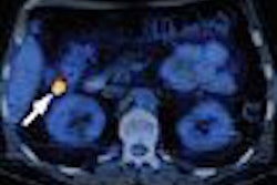
| Greater iodine concentration was a principal cause of improved image quality in two CTA scans of the same patient performed six years apart. Image (right) acquired in 2004 on a 64-slice scanner using 400 mL/kg at 5 cc/sec shows significantly better enhancement in the peripheral regions compared to the first scan (left) performed in 1998 on a four-slice scanner using 300 mL/kg iodine concentration injected at 3 cc per second. All images courtesy of Dr. Christoph Becker. |
"Because of the higher concentration of iodine in the pulmonary arteries, we see much (farther) into the periphery," he said of an image acquired with the higher-concentration Iomerol 400 agent (Bracco, Milan). The concentrated formula is approved for use in Europe, but not in the U.S.
To test the relationship between iodine concentration and flow rate, the group conducted an experiment using two different iodine concentrations, 140 mL of a 300 mgL/mL iodinated formula (Iomerol 300, Bracco) or alternatively, 140 mL of a 400 mgL/mL formula (Iomerol 400), randomized in 60 patients (63 ± 13 years, 43 male) at one of two different flow rates, 3.5 cc/sec or 2.5 cc/sec.
 |
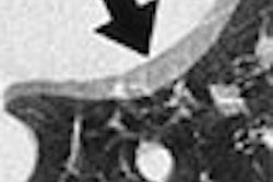
| Levocardiograms show continually improving image quality with increased CT detector rows. |
CTA images were acquired on a four-detector CT scanner (Sensation 4, Siemens Medical Solutions, Erlangen, Germany) at 120 kVp, 400 effective mAs, 1.25-mm slice thickness, and 0.7-mm reconstruction interval.
"Using the low-density contrast media with the low flow rate led most of the time to rather poor enhancement of approximately 200 HU," Becker said. When data from this protocol were evaluated, the group found that "in 50% of these cases the enhancement was not sufficient to make a diagnosis of the coronary arteries."
On the higher end of the concentration scale, the 400 mgL/mL agent combined with 3.5 cc/sec flow rate led to a rather high enhancement of approximately 350 HU in the left ventricle and the coronary arteries. The injection of approximately 1 g of iodine resulted in an optimal (250-300 HU) contrast enhancement (Investigative Radiology, November 2003, Vol. 38:11, pp. 1-5).
"But what we also saw is this: almost no difference between the high-concentration contrast media injected with low flow rates, and the low-density contrast media injected with high flow rates," Becker said.
There was also a wider range of enhancement values when the 400 mgL/mL concentration was used, compared to 300 mgL/mL. With the 400 agent, one patient might have 200 HU in the coronary arteries, the next patient 500 HU, he said.
"One of the reasons (for the difference) is that we didn't warm up the contrast media, and 400 iodine concentration has a very high viscosity," Becker said. Low temperatures may have prevented the agent from mixing thoroughly with the blood, increasing the variability of enhancement values beyond what was expected.
Recently the German team (with the assistance of Dr. Kyongtae Bae, Ph.D., from the Mallinckrodt Institute of Radiology in St. Louis), developed an equation that illustrates the predictable relationship among iodine concentration, flow rate, and enhancement. When body weight (BW) is factored in, the final enhancement of the left ventricle (HULV), can be predicted fairly accurately from the enhancement of the test bolus (HUTB) as follows: HULV = 1.13 x HUTB + 1.25 x BW.
 |
| The final enhancement of the left ventricle (HULV) can be calculated based on enhancement of the test bolus (HUTB) when body weight (BW) is factored in. |
Ultrafast 64-slice heart scans also offer the opportunity, for the first time, of targeting the contrast to specific heart regions, Becker said. The timing can be tested by first injecting a small amount of contrast followed by a saline chaser. Using this method, the contrast-enhanced CT images will at some point show contrast in the left atrium and left ventricle, but complete washout in the right atrium, right ventricle, and the coronary arteries.
This is a good start, but the method needed to be tweaked for functional analysis, which requires sharp delineation of the septal wall, Becker said. So the group designed a new protocol consisting of a main bolus of 80 cc of contrast injected at 5 cc/sec. And instead of using a pure saline chaser afterward, the saline is now mixed with the contrast media in a ratio of 1:1, Becker said.
"Right after the main bolus we're injecting 50 cc of this mixture, also injected at 5 cc/sec," he said. "You get good bright homogeneous enhancement in the left ventricular system, and you still have rather bright, but not too bright (for calcium assessment), enhancement in the right ventricular system. It allows you to delineate the septal wall and do your functional analysis."
 |
| Contrast protocol offers improved enhancement for 64-slice CTA and the ability to assess the myocardial wall. |
This method has become the group's standard 64-slice CTA contrast protocol, as it enables assessment of myocardial wall function when needed, he said.
By Eric Barnes
AuntMinnie.com staff writer
September 7, 2005
Related Reading
Part II: Free scans, general practitioners key to CTA success, July 26, 2005
Part I: Simple steps ease cardiac CTA workflow in private practice, July 8, 2005
CT nips at MRI's heels in LV function study, June 29, 2005
Multislice CT reveals previous plaque rupture in coronaries, June 14, 2005
CTA seen as accurate complement to invasive angiography, May 24, 2005
Copyright © 2005 AuntMinnie.com