Incidental focal colonic FDG uptake on PET/CT justifies performing a colonoscopy to detect malignant and premalignant lesions, French researchers have concluded. In a study of 20 patients with 21 areas of focal colonic uptake on routine PET/CT scans, the researchers found clinically significant lesions in 75% of the patients and 67% of the FDG findings. None of the patients had a history of colorectal cancer.
"Co-registered PET/CT improves the analysis of colonic FDG uptake by avoiding confusion between abnormal focal uptake and physiologic activity due to fecal stasis, smooth muscular activity, or segmental inflammation," wrote the researchers from the departments of nuclear medicine at the Rene Huguenin Cancer Research Center in Saint-Cloud, Henri Becquerel Cancer Research Center in Rouen, and Hospital Foch in Suresnes (American Journal of Roentgenology, August 2005, Vol. 185:2, pp. 495-500).
The retrospective study found that the PET/CT (Discovery LS, GE Healthcare, Chalfont St. Giles, U.K.) results were false-positive in five of the 20 patients, but 18 colonoscopic abnormalities were found in the remaining 15 patients. Colonoscopy with a pathology examination was the gold standard.
Fourteen of the 21 FDG findings on PET/CT matched colonoscopic abnormalities, which consisted of 10 adenomas, three carcinomas, and one hyperplastic polyp. The findings from colonoscopy and PET/CT were considered matching if they were present in the same segment of the colon. In one patient, the FDG finding and the polyp were not in the same site, and the finding was considered a false positive.
 |
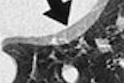
| Focal colonic FDG standardized uptake value (SUVmax = 7) was shown in cecum, with fecal stasis (arrows) on CT image. Colonoscopy was negative (patient 8). Gutman F, Alberini J-L, Wartski M, Vilain D, Le Stanc E, 3, Sarandi F, Corone1 C, Tainturier C, Pecking AP. "Incidental Colonic Focal Lesions Detected by FDG PET/CT" (AJR 2005; 185:495-500). |
Four false-negative results were found in three patients. While the positive predictive value of PET/CT to detect colonoscopic abnormalities was 61%, the false-positive rate was 33% -- seven of the FDG findings did not match any colonoscopic abnormality.
"Our results are consistent with two large studies showing FDG PET is a sensitive tool to detect premalignant lesions," the authors wrote. "Combining information from anatomic (CT) and functional (PET) image techniques improves the scan's diagnostic value compared with both techniques taken separately."
The researchers also calculated the mean standard uptake value (SUVmax) of the FDG findings. The mean SUVmax was higher in the true-positive PET/CT results than in the false-positive results (11 .2 ± 6.5 versus 7.1 ± 3.3) but the difference was not statistically significant (p = 0.14). In all, the SUVmax ranged from 3.1 to 25 with the SUVmax being 15 ± 11.6 for carcinomas and 7.1± 3.3 for negative colonoscopy.
The authors warned that proper interpretation of PET/CT scans means paying mind to possible inaccuracies of the FDG uptake location generated by peristaltic motions occurring between CT and PET image acquisitions.
Earlier studies have shown that F-18 FDG PET/CT could also play a role in detecting cancerous and precancerous lesions of the gastrointestinal tract (GIT), and that incidentally detected F-18 FDG accumulations in the GIT should be followed up with colonoscopy or other appropriate evaluation, as they lead to changes in patient management in a large number of cases.
In a study from Switzerland, researchers from the University Hospital of Zurich found that although incidentally detected lesions on F-18 FDG PET/CT (Discovery LS, GE Healthcare) occurred only in about 3% of cases in the patient group, the lesions were associated with substantial risk of an underlying cancerous or precancerous lesion. "Early identification of these occult lesions may have a major impact on the patients' management and outcome," the researchers wrote in their study in the Journal of Nuclear Medicine (November 2004, Vol. 45:11, pp. 1804-1810).
The retrospective study found cancerous lesions in 13 patients, precancerous lesions in 29, inflammatory and miscellaneous lesions in 18, and discordant findings on endoscopy in nine. Endoscopy was performed in 69 patients from the cohort of 98 patients showing incidentally detected lesions in the GIT. Follow-up findings were unavailable for the remaining 29 patients in the cohort.
 |
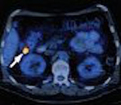
| Coronal PET/CT (A), transaxial CT (B), and transaxial PET/CT (C) images from 69-year-old man show pathologic F-18 FDG accumulation with correlative soft-tissue density (arrows) in right colonic flexure. Endoscopic and histopathologic examinations revealed advanced colonic adenoma. Ehab M. Kamel, Miriam Thumshirn, Kaspar Truninger, Marc Schiesser, Michael Fried, Barbara Padberg, Didier Schneiter, Sandro J. Stoeckli, Gustav K. von Schulthess, and Katrin D.M. Stumpe. Significance of Incidental 18-F-FDG Accumulations in the Gastrointestinal Tract in PET/CT: Correlation with Endoscopic and Histopathologic Results. J Nucl Med 2004; 45;1804-1810, Figure 2. |
Interestingly, analysis of the findings with the Fischer exact test showed 90% of the cancerous and precancerous findings and 50% of the benign findings (inflammatory and benign lesions) were characterized by focally increased F-18 FDG uptake (p = 0.001).
The study also showed that 12 of the 13 newly occurring cancer lesions occurred in those patients with primary tumors of the aerodigestive tract.
"The occurrence of 19 (70%) of 27 adnenomas among patients with aerodigestive tract strengthens our observation about the propensity of a second primary GIT tumor in subjects with a known head and neck, lung, and gastrointestinal cancer," the group stated.
The GIT findings had an impact on patient management for eight of the 13 carcinoma patients undergoing curative resection and the rest undergoing palliative chemotherapy. Among those with precancerous lesions, 27 had advanced colonic adenomas and were protected from potential malignancy by undergoing PET/CT-guided polypectomy, the authors noted.
Two patients with acute diverticulitis were treated with antibiotics and four with colitis received either conservative therapy management with follow-up colonoscopy, or an immunosuppressive therapy regimen. Overall, the GIT findings caused changes in the clinical management in 20 of the 69 patients.
"The detection of 27 advanced adenomas would have been missed, or at least postponed, if colonoscopy and excisional biopsy were not triggered by the incidental 18F-FDG PET/CT findings in our series," the group wrote.
By N. Shivapriya
AuntMinnie.com contributing writer
September 6, 2005
Related Reading
PET/CT improves diagnostic accuracy of unexpected malignancies, June 30, 2005
FDG-PET trumps CT for colon cancer staging, May 23, 2003
PET/CT boosts confidence in rectal cancer staging, US yields high sensitivity, April 3, 2003
Unexpected FDG-PET foci may signal subclinical tumors, December 26, 2002
FDG-PET signals early response of malignant GI tumors to Gleevec, July 1, 2002
Copyright © 2005 AuntMinnie.com