Radiologists in Beijing, China, have developed a joint deep learning and radiomics AI model that can flag how invasive tumors may be in patients with lung cancer, according to a study published April 9 in Radiology.
The approach could ultimately help clinicians determine which patients with suspected disease are candidates for surgery, noted led authors Zhengsong Pan and Ge Hu, PhD, of Peking Union Medical College Hospital.
“These models could assist in the preoperative care of patients with lung adenocarcinoma,” the group wrote.
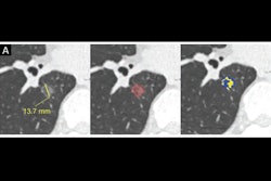
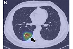
Lung adenocarcinoma is the most common primary lung cancer seen in the U.S. Tumors manifest as ground-glass nodules (GGNs) on CT scans. Deciding whether the lesions are preinvasive, minimally invasive, or invasive, however, is a significant challenge, and these determinations drive the timing of surgery, according to the authors.
To address this challenge, the group developed their deep-learning and radiomics-based approach, which classifies GGNs into preinvasive (atypical adenomatous hyperplasia or adenocarcinoma in situ), minimally invasive, or invasive adenocarcinoma.
Imaging data used in the study included CT images of a total of 4,929 nodules from 4,483 patients (mean age, 50.1 years old). The researchers divided these images into training (n = 3,384), validation (n = 579), and internal (n = 966) test sets. An external test set included a total of 361 GGNs from 281 patients.
In brief, the researchers first constructed three conventional ternary classification models: model 1 was constructed by using a radiomics method; model 2, by a deep-learning method; and model 3, by a joint learning method combining both radiomics and deep learning. The models were progressively modified through framework optimization, joint learning, and an “adjudication strategy,” the authors wrote.
Significantly, this adjudication strategy involved integrating the ternary classification models with two binary classification models to resolve discordant classifications, and this step simulates the multireader approach radiologists take to resolve discordant nodule classifications, the authors noted.
On an external validation set of 362 CT exams, the joint model combining binary and ternary classification models and adjudication of discordant classification had the highest overall performance (p < 0.001) for predicting minimally invasive adenocarcinoma (accuracy, 85%; sensitivity, 75%; specificity, 89%).
“Performance of deep-learning models for predicting preinvasive, minimally invasive, or invasive adenocarcinoma was improved by combining binary and ternary classification models,” the researchers wrote.
In an accompanying editorial, Jae Ho Sohn, MD, and Brandon Fields, MD, the University of California, San Francisco, wrote that the study ultimately confirms the importance of radiomics and deep learning in accurate risk stratification of pulmonary adenocarcinoma spectrum lesions.
Future research directions may include a more granular assessment of the radiomics features that were most predictive of invasiveness, they noted.
“[This] study will serve as a valuable stepping stone to constructing a multimodal approach when determining the most appropriate candidates for surgery in patients with suspected pulmonary adenocarcinoma lesions,” Sohn and Fields concluded.
The full study is available here.