Have you ever diagnosed a three-foot-tall 3D heart? Talk about cardiomegaly. Researchers from the Netherlands who survived the experience found they could diagnose valve defects that went unnoticed in a standard 2D exam.
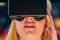
"3D images are used increasingly nowadays to visualize complex pathology of the heart as opposed to the more traditional 2D approach," said Anton Koning, Ph.D. "However, these nice 3D images are still generally only presented on 2D media, either a flat screen or a printout. We think the dignosis with ultrasound can be improved by using steroscopic displays in different kinds of VR (virtual reality) systems, and that’s why we’re conducting this research in 3D ultrasound."
The researchers at Erasmus University Medical Center in Rotterdam wanted to know, first, if the I-Space VR system (Barco, Kortrijk, Belgium) was feasible for 3D ecocardiography, and second, if it offered any additional information, said Koning, who is a research scientist at Erasmus University’s bioinformatics department.
The researchers felt that information critical to the diagnosis could be lost on a standard 2D display, Koning said at the 2007 Computer Assisted Radiology and Surgery (CARS) meeting in Berlin.
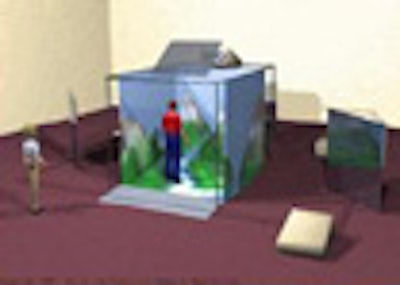
For their 3D studies the researchers used a Barco CAVE (Computer Automatic Virtual Environment) virtual reality system. The CAVE I-Space immerses several viewers in a 3D virtual world, Konig said. Conceived in the early 1990s as the successor to the head-mounted display (i.e., the VR helmet), early versions of the CAVE housed only a single reader, "which precludes successful collaboration with colleagues," he said.
The hospital installed a newer version of the CAVE in 2004. It projects images stereoscopically on the walls, floor, and/or ceiling of a small room, which typically ranges in size from 2.5 x 2.5 x 2 m to 4 x 4 x 3 m. "As we manipulate the data we have an arbitrary cutting plane, and also free hand to cut away or erase certain parts of the data," Koning said.
Projection around the viewers allows easy collaboration and prevents so-called "simulation sickness," Koning said. High-resolution projection allows visualization of fine details, enabling large amounts of information to be displayed simultaneously.
 |
| The Barco CAVE (Computer Automatic Virtual Environment) VR system installed at Erasmus Medical Center is equipped with eight DLP (digital light processing) projectors (1280 x 1024, 60 Hz, 1500 lumen, 1300:1 contrast ratio) projecting images in passive (visual) stereo based on circular polarization. Projectors are housed in three rigid walls plus a floor. The rectangular screens measure 2.60 x 2.08 meters. So-called passive stereo provides separate images for the left and right eye of the viewer. There are filters in front of projectors and in glasses worn by viewers, who can collaborate on the diagnosis. All images courtesy of Anton Koning, Ph.D. |
 |
| The Barco CAVE I-Space installation at Erasmus includes wireless tracking, 3D joysticks with four buttons, and a collaboration option (eyepoint/input switching). The SGI Prism (Silicon Graphics, Sunnyvale, CA) image generator includes eight Itanium2 processors, 12 GB shared memory, and eight dual-head high-end graphics cards. |
 |
| The Barco CAVORE (Cave Volume Renderer) system provides volume rendering of medical and other datasets, true 3D views of data at any scale, high-resolution, high-performance rendering, and real-time, interactive manipulation. Features include an arbitrary cutting plane and free-hand eraser, gray value, color and transparency transfer functions for segmentation, and distance and volume measurements. |
The researchers have used the CAVE to experiment with multimodality imaging, fetal imaging, and other applications. Recently Koning and his colleagues (G. Bol Raap, A.E. van den Bosch, F.J. Meijboom, A.J.J.C. Bogers, P.J. van der Spek) performed two small studies to test radiologists’ ability to visualize cardiac pathology using the system.
 |
| In addition to echocardiography, Erasmus researchers have used the I-Space system for whole-body CT, 3D ultrasound and MRI fusion, and prenatal ultrasound. |
Study one
The first study included 3D echocardiographic datasets from two normal subjects and four patients with mitral valve pathology.
Ten observers, including five cardiologists, three cardiologists in training, and two cardiothoracic surgeons, all without prior knowledge of the subjects, were asked to assess both the mitral valve anatomy and any pathology.
The researchers had to convert the echo data, scanned on a Sonos 7500 scanner (Philips Medical Systems, Best, Netherlands), into a format readable by the CAVORE application, but this step took less than a minute per case, Koning said.
After 10 minutes of instruction time in the I-Space, all observers were able to use the virtual pointer to manipulate the volume and make the necessary cut planes, Koning said. All observers correctly assessed the mitral valve morphology in all holograms, with an average analysis time of approximately 10 minutes per study.
 |
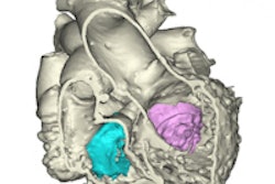
| Mitral valve specimen (left) and 3D echo images (right) seen from left atrium (top) and left ventrical (bottom). |
Study two
"For the second study we used 12 3D interoperative echo datasets of patients undergoing suture closure of a ventricular septal defect -- a hole in the heart," he said.
The data were evaluated in real-time 2D mode during the operation, and postoperatively using 3D views in the I-Space. "The aim was to assess whether tricuspid valve leaflet mobility was affected," Koning said.
All patients were able to be evaluated in 3D, he said. Intraoperative 2D assessment showed no valve stenosis or regurgitation, and normal leaflet mobility. However, 3D analysis in the I-Space revealed three patients with restricted mobility of the septal tricuspid valve leaflet.
"This means that in three of five patients, the 2D diagnosis was incorrect," Koning said. Tempering their success, in four 3D datasets the posterior leaflet and papillary apparatus were only partially included, meaning that 3D analysis wasn't ideally suited for these four patients, he added.
 |
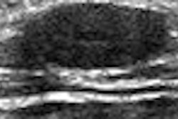
| Echocardiography exams are viewed in 3D in the CAVE. The flat appearance of the photograph is deceptive, according to Koning; observers see the anatomy in 3D. |
"We can certainly say that the use of VR of dynamic echocardiography data is feasible in the I-Space, and we demonstrated its usefulness," Koning said. "Also, we found that (when) using a normal (2D) workstation, the learning curve is much steeper. We were able to use (I-Space) in only 10 minutes -- a normal (2D) workstation requires a multiday course." Diagnostic time is about the same as a 2D workstation, at 10 minutes per case.
Still, the gargantuan workstation has been a difficult fit for the cardiology department, at least in its present form. "For echocardiography the full CAVE system is a bit of an overkill," Koning said. "In general we want to have the equipment as close as possible to the desk."
Future goals including adapting the installation to run on smaller VR equipment, either based on a single projection surface or a CRT or LCD monitor, he said. The group is currently conducting a study of ultrasound fetal measurements in 40 pregnant women, Koning told AuntMinnie.com.
By Eric BarnesAuntMinnie.com staff writer
August 2, 2007
Related Reading
Virtual human puts doctors inside their patients, May 24, 2007
Image-guided surgery market in U.S. to reach $300 million, January 16, 2007
Advanced visualization alone reduces coronary CTA accuracy, December 22, 2006
Part III: Image processing has room to grow, October 23, 2006
3D lab efficiency may depend on who's in control, June 22, 2006
Copyright © 2007 AuntMinnie.com