Thorax 2000 Feb;55(2):143-6
Adult familial cryptogenic fibrosing alveolitis in the United Kingdom.
Marshall RP, Puddicombe A, Cookson WO, Laurent GJ.
BACKGROUND: Familial cases of cryptogenic fibrosing alveolitis (CFA) have
previously been reported; however, the prevalence and genetic background of this
disorder are not known. The clinical and epidemiological findings of 25 families
identified within the UK are reported. METHODS: Adult pulmonary physicians in
the UK were asked to identify all families under their care in which two or more
individuals had been diagnosed with fibrosing alveolitis of unknown cause. A
detailed structured questionnaire was sent to each proband to delineate possible
environmental/occupational exposures and to obtain complete pedigree data.
Physicians were also asked to provide clinical and diagnostic information.
RESULTS: Twenty five families were identified comprising 67 cases. Suitable data
for analysis were available for 21 families (57 cases). The male:female ratio
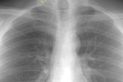
was 1. 75:1 (p<0.05). A high resolution computed tomographic (HRCT) scan was
performed in 93% and a diagnosis of CFA confirmed on biopsy specimens in 32%.
The mean age at diagnosis was 55.5 (2.5) years. Fifty percent of cases were ever
smokers and 18% had been diagnosed as asthmatic. Exposure to known fibrogenic
agents was recorded by 36% of patients. Clinical signs/symptoms and histological
findings were indistinguishable from non-familial cases. CONCLUSIONS: This study
represents the largest cohort of familial CFA cases reported to date and
confirms a prevalence of 1.34 cases per 10(6) in the UK population. Although
rare, such cases represent an important subgroup in which a genetic
susceptibility to pulmonary fibrosis is particularly evident. Familial patients
are younger at diagnosis but otherwise indistinguishable from non-familial
cases. The mode of inheritance is as yet unclear but a number of genetic loci
are likely to be involved and are the subject of ongoing studies.