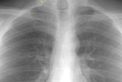
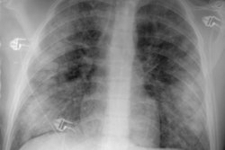
Usual Interstitial Pneumonitis:
The PA and lateral chest radiographs in this patient demonstrate the classic findings in UIP. The lung volumes are diminished. There is a coarsened prominence to the interstitial markings predominantly in a basilar and posterior distribution (noted best on lateral exam), and the heart border has a shaggy appearance (click images to enlarge).

The CT scan (supine and prone images) reveal extensive interstitial fibrosis with honeycombing.