(Ultrasound Review) Plantar fibromatosis can be evaluated with sonography according to researchers at Prince of Wales Hospital in Hong Kong. Their study described the ultrasound appearances of plantar fibromatosis and was published in the November issue of American Journal of Roentgenology.
According to the authors, this disease is characterized by proliferation of fibrous tissue within the plantar fascia usually in the mid- and forefoot area, and patients most often present with either foot pain or a lump in the sole of the foot.
In this retrospective study of 14 patients, they reviewed the clinical presentation, sonographic appearances, and clinical progress over a three-year period. Ultrasound imaging of the plantar fascia of both feet was performed using high-frequency linear array transducers.
Patients were scanned in a prone position with the feet resting over the end of the table. The thickness of the plantar fascia at the calcaneal insertion was measured, and a thickness of 4.5 mm or more was thought to be abnormal. The ultrasound characteristics assessed included location, size, and appearance of plantar fibromatosis nodules. These characteristics then were compared to symptom duration and clinical outcome.
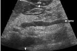
They discovered 25 fibromatosis nodules in 19 feet. The nodules appeared as "discrete fusiform nodular thickening of the plantar fascia, separate from the calcaneal insertion." Sixty percent of lesions were located medially, and 40% were centrally placed in the fascia. They found that most nodules were hypoechoic (76%), and well defined (64%), without acoustic enhancement (80%).
"One quarter of the affected feet had coexistent thickening of the plantar fascia at the calcaneal insertion with no related symptoms," they said.
Problems affecting the sole of the foot can be divided into fascial and non-fascial pathologies. Ultrasound demonstrated the continuity between the lesion and the plantar fascia, which eliminated other lesions such as neuroma or extrafascial soft tissue tumors including "ganglion, inclusion cyst, foreign body granuloma, nerve sheath tumor, or synovial sarcoma."
The authors described the two main differential diagnoses of plantar fibromatosis: plantar fasciitis and chronic rupture of the plantar fascia.
"Plantar fasciitis is seen sonographically as a thickening and hypoechogenicity of the plantar fascia at or near the calcaneal insertion, especially medially, and is often associated with a calcaneal spur."
In contrast, plantar fibromatosis occurs in the plantar fascia, separate from the calcaneum. They admitted that differentiating plantar fibromatosis from a chronic partial rupture of the plantar fascia could be more challenging, as both appear as a fusiform plantar fascial mass separated from the calcaneal insertion on ultrasound. They said that acute or repetitive trauma, and a visible tear or perifascial edema or fluid on ultrasound characterized chronic partial rupture.
None of the patients studied were surgically treated, and so they were unable to compare the ultrasound and histologic features. They reported, "although the lesions in this study varied in size, definition, and echogenicity, no relationship between any of these features and the chronicity of symptoms was found."
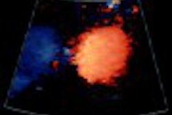
Color Doppler was used to assess vascularity, but only two nodules showed distinct vascularity and this was not associated with any particular clinical feature. Although MRI is an excellent means of diagnosing plantar fascia nodules and disease extension, ultrasound provides a low cost alternative that is more readily available, they said.
The authors concluded, "the sonographic appearances of plantar fibromatosis vary but are still characteristic enough to allow a specific diagnosis to be made." However, they found no relationship between the ultrasound characteristics of the nodules and patient outcome.
Sonography of plantar fibromatosisGriffith, J F et al
Department of diagnostic radiology and organ imaging, Chinese University of Hong Kong, Prince of Wales Hospital, Shatin, New Town, Hong Kong
AJR 2002 November; 179:1167-1172
By Ultrasound Review
December 17, 2002
Copyright © 2002 AuntMinnie.com