New techniques such as 3-D ultrasound, ultrasound contrast agents, and B-flow imaging offer tremendous potential in aiding vascular diagnosis, according to Dr. John Pellerito, chief of ultrasound, body CT, and MRI at North Shore University Hospital in Manhasset, NY.
"These techniques are complementary and work very well together in powerful combinations to improve diagnosis," he said. "And there's also the potential for new applications as well."
Pellerito discussed these techniques at the 2002 Leading Edge in Diagnostic Ultrasound conference in Atlantic City, NJ.
Of course, any discussion of vascular ultrasound must encompass power Doppler, which remains an integral part of current vascular ultrasound practice, and is a complementary tool for the new techniques, Pellerito said.
"I use (power Doppler) in multiple vascular applications and find it complementary to color and pulsed Doppler," he said.
Ultrasound contrast
Contrast agents show promise in vascular ultrasound, owing in part to their potential to increase the number of diagnostic studies and improve cost-effectiveness by decreasing sonographer and physician time, and reducing the need for additional imaging procedures, Pellerito said. In addition, ultrasound contrast may open the door for new vascular ultrasound applications.
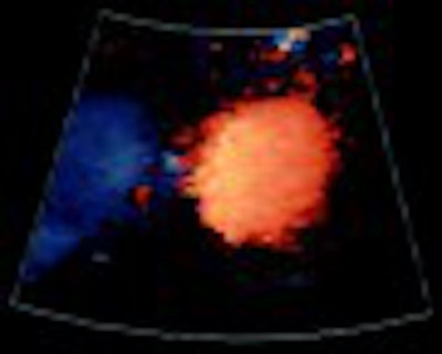
 |
| Precontrast color Doppler image with limited visualization of the right renal artery. |
According to Pellerito, potential applications for contrast agents include:
- Transcranial Doppler studies.
- Studies for the differentiation of severe carotid artery stenosis from occlusion.
- Peripheral vascular studies.
- Studies for the assessment of bypass graft patency and venous thrombosis.
Ultrasound contrast may also be suitable for evaluating peripheral arteries, bypass grafts, and renal and mesenteric arteries, he added.
In addition, the agents can aid in characterizing tumors, such as identifying malignant neovascularity in indeterminate liver, kidney, and breast lesions. The ability to evaluate changes in tissue perfusion can identify gonadal torsion, organ infarction, and inflammation. Detection of inferior vena cava and portal vein thrombosis may be improved with ultrasound contrast. The evaluation of TIPS (transjugular intrahepatic portosystemic shunts) may also benefit, Pellerito said.
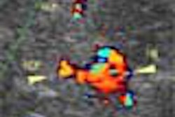
 |
| Following administration of ultrasound contrast, a long segmental stenosis with a beaded appearance (arrow) is seen consistent with fibromuscular dysplasia. |
Ultrasound contrast efficacy can also be bolstered by power Doppler and harmonic imaging techniques, he said. In preliminary studies, contrast-enhanced power Doppler has yielded improved blood-flow visualization, although flash (clutter) artifacts may be present. Adding harmonic-imaging techniques such as second harmonic and pulse inversion can reduce clutter artifacts, Pellerito said.
Harmonic imaging can provide improved demonstration of normal anatomy and vascular disease, and can also assist in the identification and characterization of solid tumors. It's also a valuable adjunct to 3-D ultrasound, Pellerito said.
Three-dimensional US
Much of the research in 3-D ultrasound has revolved around obstetric, gynecological, and cardiac applications, but the technology offers potential in vascular applications as well, Pellerito said. Although ultrasound 3-D capabilities currently lag behind those available in MR and CT, that level of technology will ultimately reach ultrasound as well.
"If you've seen some of the stuff you get with CT and MR workstations today, it really blows you away," he said. "Not only do you get volume, but you can just look at vessels and soft tissue, and take out the bone. Ultrasound is not there yet; it will be a couple of years before we have that accessibility to data. But when it comes, it's going to be very important and we're all going to be using it."
Three-dimensional imaging offers the ability to obtain anatomical views not possible on 2-D imaging, he said. Users can reformat the image volume in different imaging planes, extract information obscured by tissue or artifacts, and perform a surface or transparent display. In addition, 3-D ultrasound allows offline measurements to be made of regions of interest not noted on the initial exam and to obtain new additional information, Pellerito said.
Pellerito said that recent research has shown utility for 3-D exams of the carotid arteries and cerebral circulation. Other potential vascular applications include cardiac imaging, renal and liver vessel mapping, and visualization of tumor vascularity for lesion characterization, he said.
B-flow
A new technique already impacting vascular imaging is B-flow, a B-mode imaging technique that enables simultaneous, direct gray-scale visualization of blood echoes and tissue, Pellerito said. With B-flow, digitally encoded wide-band pulses are transmitted for each scan line, and the returning signals are decoded and filtered to provide increased sensitivity to moving blood cells, and distinguish blood from tissue in real-time, he said.
This technique offers improved resolution, and is helpful when visualizing abnormal flow profiles, Pellerito said. In comparison with color Doppler, B-flow imaging is characterized by less averaging, flash artifact, and overwriting.
"And I think there's improved boundary detection (with B-flow)," he said.
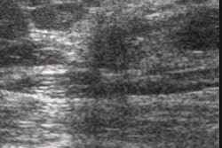
 |
| B-flow image provides improved definition of the vessel lumen (straight arrow) and demonstrates ulceration, which was not detected on the Doppler study but confirmed at arteriography. Images courtesy of Dr. John Pellerito. |
B-flow benefits include improved carotid plaque characterization and better demonstration of arterial stenosis and turbulent flow states. B-flow also offers potential in evaluation of high-flow lesions in dialysis fistula and bypass grafts, where color-flow artifacts can obscure the underlying stenosis.
B-flow can demonstrate venous thrombosis, as well as small clots that can be distinguished from flowing blood, Pellerito said. Further research is needed to determine whether B-flow will aid in the diagnosis of diseases of the aorta and deeper branch vessels, he said.
By Erik L. RidleyAuntMinnie.com staff writer
July 8, 2002
Related Reading
IVUS after angiography pinpoints chronic PE, February 4, 2002
Vascular Diagnosis with Ultrasound, January 29, 2002
Imaging explores link between kidney failure and atherosclerosis, January 28, 2002
New imaging techniques spark cardiovascular diagnostics growth, September 19, 2001
Transcranial Doppler an insensitive measure of cerebrovascular reserve, August 24, 2001
Copyright © 2002 AuntMinnie.com