Acquisition of PET images as long as six hours after FDG injection can be a useful and easy-to-perform technique in delineating gliomas from gray matter and detecting cerebral lymphomas.
The finding comes from research based at the department of nuclear medicine and radiology at Jean Minjoz University Hospital in Besançon, France, and presented at the 2007 RSNA meeting in Chicago.
In detailing results of the study, Dr. Oleg Blagosklonov noted that the very delayed (after six hours) cerebral FDG-PET scan can reveal both hypermetabolic and hypometabolic lesions in oncological and nononcological patients. The research team's analysis extends the value of delayed or dual-time-point (two to three hours after injection) FDG-PET imaging for detecting abnormal lesions.
All the PET scans reviewed in the study were conducted in Jean Minjoz's nuclear medicine and radiology department. From January 2005 to March 2007, 58 cerebral FDG-PET scans were performed in 44 patients with the following diagnoses:
- Cerebral metastasis suspected on MRI (15)
- Glioma (12)
- Pituitary adenoma or Cushing's syndrome (6)
- Epilepsy (4)
- Cerebral lymphoma (2)
- Unknown (5)
After six hours of fasting, 4-5 MBq/kg of FDG was administered to patients intravenously. Then PET images were acquired twice, with the first scan one hour after injection and the second scan six hours after injection, using a Biograph Duo PET/CT system (Siemens Medical Solutions, Erlangen, Germany).
The CT portion of the procedure was performed in a spiral mode with 2-mm slice thickness. FDG images were acquired with an axial field-of-view of 12 cm.
 |
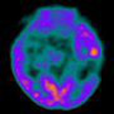
| Images show high-grade transformation of an oligodendroglioma visualized at the delayed PET scan. All images courtesy of Dr. Oleg Blagosklonov and Jean Minjoz University Hospital. |
A standard one-hour PET scan showed no lesion in one case, one or more high-uptake lesions in 18 cases, and one or more low-uptake lesions in 39 cases, Blagosklonov noted. The delayed acquisition revealed an additional 11 high-uptake lesions, allowing the detection of six recurrent cerebral metastases, three high-grade gliomas, one pituitary microadenoma, and one focus of epilepsy.
 |
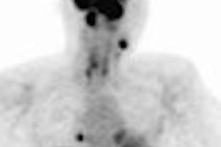
| Researchers observed enhanced contrast between gray matter and all hypermetabolic lesions, even visible at the first PET scan. Above, a cerebral metastasis. |
 |
| Contrast between gray matter and hypometabolic lesion also was enhanced by the very late acquisition technique. The image shows a low-grade glioma. |
Blagosklonov said the research shows that the easy-to-perform and well-accepted technique is comparable to FDG-PET scanning without attenuation correction in lung cancer diagnosis.
By Wayne Forrest
AuntMinnie.com staff writer
December 21, 2007
Related Reading
PET does not predict survival in advanced lung cancer, November 29, 2007
FDG-PET/CT accurately detects inflammatory breast cancer, November 27, 2007
Lack of IV contrast causes discrepancy in CT with PET scans, November 22, 2007
FDG-PET works for nose cancer staging, study finds, November 15, 2007
Dual-time-point PET spots local, distant breast metastases, September 14, 2006
Copyright © 2007 AuntMinnie.com