NEW ORLEANS - The Society of Nuclear Medicine chose an image produced by a lutetium oxyorthosilicate (LSO) PET/CT whole-body scan as its 2003 Image of the Year. The award was selected and announced by Dr. Henry Wagner, director of Radiation Health Sciences at Johns Hopkins in Baltimore.
The poster study behind the image, LSO-PET/CT Whole Body Imaging in 7 Minutes: Is It Feasible?, was conducted by Dr. Benjamin Halpern and colleagues from the University of California, Los Angeles.
The group devised a weight-based PET image acquisition protocol for clinical PET/CT studies that enables whole-body scans to be conducted in 7 to 28 minutes. The studies were performed on a CPS Innovations of Knoxville, TN, (a joint venture between Siemens Medical Solutions of Malvern, PA, and CTI Molecular Imaging, also of Knoxville) LSO PET/dual-slice CT unit at the Ahmanson Clinic for Biological Imaging at the UCLA School of Medicine.
"One of the major criteria that I use in selecting the image of the year is its potential significance in the field of nuclear medicine," Wagner said. "It is my belief that one of the things we have to do in nuclear medicine is to extend it to as many patients as possible; I believe that we are moving into a high-throughput world. We don't want to have PET and SPECT and other technologies of nuclear medicine become so rate-limiting that they're bypassed."
 |
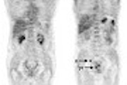
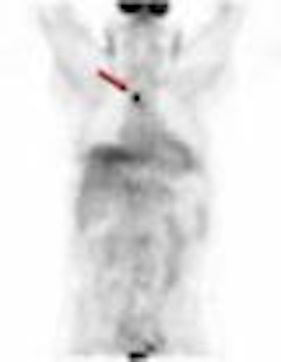
| A 60-year-old patient with recurrent head and neck cancer and body weight of 225 lb. (102 kg.) presents with a 1.5 cm superior mediastinal lymph node cancer (red arrow) in an image obtained at three minutes during an upper-body PET scan. Image courtesy of the Society of Nuclear Medicine and the Ahmanson Clinic for Biological Imaging at the UCLA School of Medicine. |
According to a member of the study team, Dr. Johannes Czernin, the facility has used the weight-based PET protocol to conduct approximately 1,200 whole-body scans at about 15 minutes per scan, and has discovered 120 lesions as a result.
The researchers prospectively compared PET image quality and lesion detectability using 1-, 2-, 3-, and 4-minute PET images obtained in the same patient in a total of 32 patients. Each patient was injected with 0.21 mCi/kg of FDG and underwent a whole-body standard CT scan (45 seconds). PET emission data was acquired at 4 minutes per bed position for 24 patients, and at 3 minutes per bed position for 8 patients.
"From the 3- or 4-minute scans, 1-minute frames were extracted in order to reconstruct 1-to-4-minute scans for each patient," the researchers wrote.
The team then divided the patients into three weight groups: those weighing less than 130 lbs (59 kg); those who weighed between 130-179 lbs (59-82 kg); and those weighing between 180 and 249 lbs (82-113 kg). Three independent observers then rated image quality as good, moderate, poor, or nondiagnostic in quality, recording the number and location of hypermetabolic lesions.
The group found that for patients weighing less than 130 lbs (59 kg), the 1-minute scans per bed position provided the same diagnostic information as the 2-, 3-, and 4- minute scans. For the subgroup of patients weighing between 130 and 179 lbs (59-82 kg), the 2-minute scans provided the same diagnostic information as the 3- and 4- minute scans. For the largest group of patients, who weighed between 180 and 249 lbs (82-113 kg), the 3-minute scans provided the same diagnostic information as the 4-minute scans.
Still, "Image quality improved with scan duration," the authors wrote.
The scans were interpreted as concordantly positive or negative in 73% of all patients. The researchers stated that concordance rates were 89%, 68%, and 69% for each of the weight groups, respectively, and that imaging time did not affect concordance rates and lesion detectability in any of the weight groups.
"While image quality can be improved by longer acquisition times, diagnostic information is not compromised with fast PET protocols," the group concluded. "The approach allows for considerable time savings and may decrease artifacts associated with patient motion."
By Jonathan S. BatchelorAuntMinnie.com staff writer
June 24, 2003
Related Reading
PET, SPECT studies chosen as SNM images of the year, June 18, 2002
Wagner taps PET Alzheimer?s study as SNM 2001?s Image of the Year, June 26, 2001
Copyright © 2003 AuntMinnie.com