Although MRI is commonly used for evaluating multiple sclerosis (MS), it is anything but an ideal imaging exam. Traditional T2-weighted images do not consistently correlate with the clinical manifestations of disease extent or lesion activity, and MS symptoms vary depending on the location and pathology of those lesions.
Two recent studies have used more advanced techniques to work around MRI's shortcomings in MS imaging. In an experimental study, a group from Robarts Research Institute in Ontario, Canada, tracked cellular neuroinflammation with MR microscopy (MRM) and a superparamagnetic iron oxide (SPIO) contrast agent. This noninvasive method could reveal important markers for early MS.
And in a retrospective patient review, neuroradiologists at Duke University in Durham, NC, used diffusion tensor MRI to redefine MS plaques on fractional anisotropy (FA) maps. The group's results may offer a more accurate way to track the true extent of disease burden.
Macrophage infiltration
Macrophages are involved in every stage of MS disease progression, from inflammation to blood-brain barrier damage to demyelination. Dr. Paula Foster and colleagues from Robarts' Imaging Research Labs looked to nondestructively define macrophage infiltration in experimental autoimmune encephalomyelitis (EAE), which is an animal model of MS.
For this study, EAE was introduced in female rats that subsequently underwent whole-brain imaging on a 1.5-tesla scanner (CVi/MR, GE Healthcare, Waukesha, WI). T1-weighted multislice spin-echo images were acquired pre- and postinjection of 100 mmol/kg of Magnevist (Schering AG, Berlin).
Rat brain specimens were imaged on the same scanner, outfitted with a custom-built gradient-insert radiofrequency coil and a 3D fully refocused gradient-echo sequence, 3D FIESTA, also known as true-FISP. The SPIO agent (Feridex, Berlex Imaging, Wayne, NJ) was injected into seven rats at a rate of 22.4 mg Fe/kg.
"Intravenously administered iron oxides, taken up by macrophages in the brains of EAE animals, could be visualized with MRI. This approach has been termed 'active labeling' of cells," the group explained. "SPIO (was) administered slowly during inflammation in EAE" (Molecular Imaging, April 2004, Vol. 3:4, pp. 1-11).
According to the results, gadolinium-enhanced in vivo imaging indicated blood-brain barrier permeability in 13 EAE rats. In the brain specimens, small, localized regions of signal void (RSV) were visualized in all the rats injected with SPIO. The investigators noted that using the FIESTA sequence resulted in increased signal-to-noise ratio (SNR) and contrast-to-noise ratio, allowing for the enhanced detection of RSV.
"These small abnormalities may represent the early cellular abnormalities and events in lesion formation that have not been visualized previously in similar EAE studies," they said. "The FIESTA sequence provides substantially enhanced SNR ... as a result of the multiple signal refocusing paths available for tissues."
The next step will be in vivo imaging of EAE in the rat brain, the authors concluded. Ultimately, being able to pinpoint clinical markers in MS will help guide treatment, as well as response to anti-inflammatory therapies.
Sizing up MS plaques
Diffusion tensor imaging has proven adept at assessing the microarchitecture of the brain. In addition, fractional anisotropy (FA) values -- the degree to which water molecules diffuse in one direction -- for normal-appearing white matter are lower when adjacent to MS plaques, according to a study by Dr. Susan Kealey and co-authors in the American Journal of Roentgenology. FA maps offer good gray-white matter contrast and a high contrast-to-noise ratio.
Kealey's group set out to compare the size of MS plaques with lesion size as defined by a thresholding technique on FA maps. They suggested that combining FA maps and diffusion tensor imaging would produce substantial differences in plaque size compared to conventional MR.
 |
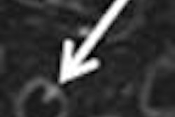
| T2-weighted images of 29-year-old woman illustrate centrifugal method of interrogating normal-appearing white matter (WM) adjacent to MS plaques. Axial T2-weighted MR image shows demyelinating plaque adjacent to anterior horn of left lateral ventricle. Kealey SM, Kim Y, Provenzale JM, "Redefinition of Multiple Sclerosis Plaque Size Using Diffusion Tensor MRI," (AJR 2004;183: 497-503). |
The study population consisted of 20 patients who underwent conventional MR scans (T2-weighted, with and without Magnevist contrast). Diffusion tensor MR images of the entire brain were acquired on a 1.5-tesla MR scanner (Signa, GE Healthcare). Images were obtained in six orthogonal directions using a standard head coil.
"Our criteria for deciding whether a lesion on T2-weighted images represented an MS plaque were that it be oval in appearance, oriented toward or abutting the lateral ventricles, and lacking associated restricted diffusion," they wrote (AJR, August 2004, Vol. 183:3, pp. 497-503).
A threshold of 40% decrease in FA was set for defining abnormal-appearing white matter on FA maps.
 |
| In the same patient, initial image of normal-appearing WM adjacent to this MS plaque, three regions of interest reached 40% fractional anisotropy (FA) reduction threshold and were recorded. Kealey SM, Kim Y, Provenzale JM, "Redefinition of Multiple Sclerosis Plaque Size Using Diffusion Tensor MRI," (AJR 2004;183: 497-503). |
In all, 36 white-matter plaques were examined in the 20 patients. On conventional MRI, the mean plaque area was 60 mm2. The mean plaque FA was 0.251; the mean FA of normal white matter was 0.429 for normal white matter. Applying the 40% FA reduction threshold, the mean combined area of abnormal white matter was 87 mm2 or 145% of the mean plaque area as seen on T2-weighted imaging.
 |
| Final area of abnormal-appearing WM is shown that comprises original plaque and four regions of interest reaching 40% FA reduction threshold. Kealey SM, Kim Y, Provenzale JM, "Redefinition of Multiple Sclerosis Plaque Size Using Diffusion Tensor MRI," (AJR 2004;183: 497-503). |
"These values suggest that current MRI sequences underestimate the amount of diseased white WM in MS ... both myelin loss and direct disruption of the nerve axons would result in great random motion of water molecules ... resulting in reduced FA values in areas of damaged tissue," they wrote.
This study's conclusions may be particularly important for MS pharmacology trials that rely on T2-weighted MRI for gauging therapeutic effect, the authors stated. Using diffusion tensor imaging and FA values instead of traditional MRI may provide new information for optimizing clinical treatment.
By Shalmali Pal
AuntMinnie.com staff writer
August 11, 2004
Related Reading
MS patients on high-dose beta interferon should stick to regimen, July 22, 2004
Multiple sclerosis drug enhances brain motor activity, June 14, 2004
Statin reduces MRI lesions of MS in pilot study, May 14, 2004
MR spectroscopy may predict disease progression in MS, December 8, 2003
Copyright © 2004 AuntMinnie.com