Previous reports have shown that FDG-PET complements traditional breast cancer imaging modalities with regard to staging, metastatic or recurrent disease, and treatment planning. But does it follow that all breast cancer patients benefit from the information offered by PET scans?
Not necessarily, according to Dr. William Eubank and colleagues, who performed a retrospective analysis of the impact of FDG-PET on patients with breast cancer. Specifically, they assessed the referral patterns for patients, as well as how valuable the modality was for defining the extent of disease.
But first, a group from New York City, lead by Dr. A. Jill Leibman, surveyed the current use and efficacy of breast PET, revealing which specialty (besides imaging) was most sold on the modality.
Who picks PET?
"The purpose of this study was twofold: One was to investigate the clinical efficacy of PET in patients with known breast cancer," said Leibman during a presentation at the 2004 American Roentgen Ray Society (ARRS) meeting in Miami Beach, FL. "The other was to determine the current utilization patterns of PET amongst our referring clinicians." Leibman is with St. Luke's-Roosevelt Hospital Center and Columbia University College of Physicians and Surgeons. Her co-author, Dr. Muni Ghesami, is from St. Luke's.
The duo retrospectively reviewed all patients who were referred for breast PET over a 10-month period. In total, 18 patients were identified and their clinical histories, and relevant imaging studies, were then reviewed.
In a dozen cases, the referring physician was a medical oncologist; in four cases, a breast surgeon. One patient was referred by a radiation oncologist and one other by a general physician, Leibman said. Prior to the PET exams, imaging work-up included bone scans, MRI, and CT, she added.
According to the study, PET results were positive in nine patients. PET upstaged disease in three out of five patients. In general, PET closely correlated with the initial impressions of the extent of disease given by the referring clinicians, she reported.
However, out of the nine, three had findings that were discordant with another imaging study. In one case, PET results were negative, while a bone scan and a CT scan were positive.
"CT demonstrated (possible) metastatic disease in the lumbar spine. A bone scan was suspicious for skeletal metastatic disease. Our initial PET study did not show any evidence of metastatic disease in the spine or the pelvis," Leibman explained. "A few months later we repeated the PET scan and we found that, at that point, there were extensive skeletal metastases. It may be necessary to repeat a PET scan several months after a positive bone scan just to make sure that the findings are concordant or discordant."
As with past studies, the group also found that PET was particularly useful in determining distant metastases and recurrent disease, which may explain why it is particularly popular with medical oncologists. In addition, a negative PET increased the specificity of bone scan findings. Unlike CT, PET can reliably distinguish post-therapeutic changes and is not influenced by body habitus, contrast bolus, or artifacts.
Overall, "PET can reduce the number of imaging evaluations of breast cancer patients," Liebman concluded.
FDG-PET for advanced cancer
In another study, Eubank's group also analyzed referral patterns for FDG-PET and evaluated the impact of the modality on defining the disease. They then went a step further and hypothesized that the impact of PET in the therapeutic plan may be greater in subgroups of patients.
"A distinguishing feature of the management of breast cancer is the need for a multidisciplinary approach … any strategies and agents offer patients with advanced breast cancer therapeutic benefits … choosing the most appropriate therapy depends primarily on accurately defining the extent of disease," wrote Eubank's group in the American Journal of Roentgenology (August 2004, Vol. 183:2, pp. 479-486).
Lead author Eubank is from the Puget Sound Veterans Affairs Health Care System in Seattle. His co-authors are from the divisions of nuclear medicine and medical oncology, and the departments of radiology and radiation oncology at the University of Washington, also in Seattle.
This retrospective study reviewed the records of 125 consecutive breast cancer patients referred for PET imaging. The primary neoplasm in a majority of the women (66%) was infiltrating ductal carcinoma. In addition to PET exams, the patients underwent conventional imaging with CT, MRI, or bone scans.
 |
| Fifty-four-year-old woman with axillary node-positive infiltrating lobular carcinoma of the right breast. Patient had been disease-free on systemic hormonal therapy until six years after initial diagnosis, when tumor markers became elevated. Posterior projection of restaging bone scan shows multiple foci of radiotracer uptake in spine, pelvis, and right rib, consistent with metastatic disease. Eubank W, Mankoff D, Bhattacharya M, Gralow J, Linden H, Ellis G, Lindsley S, Austin-Seymour M, Livingston R, "Impact of FDG-PET on Defining the Extent of Disease and on the Treatment of Patients with Recurrent or Metastatic Breast Cancer," (AJR 2004; 183: 479-486). |
PET's effectiveness for determining disease was categorized as either increased, decreased, or no change. The impact of PET on the management plan was placed into one of three categories: altered, supported, or no change.
Whole-body PET imaging was done on an Advance scanner (GE Healthcare, Waukesha, WI), 45 minutes after the IV injection of 244-400 MBq of FDG. Results were confirmed by either histopathology or follow-up imaging.
The majority of these patients were referred for PET for either the evaluation of disease response, or viability after therapy (35%). Overall, the authors reported that FDG-PET results were positive at one or more sites in 94 patients (75%) and negative in 26 (21%).
Compared to conventional imaging, PET results enabled radiologists to upstage the extent of disease in 43% of the patients, downstage it in 24%, and rendered no change in 33%. The results altered the therapeutic plan in 32% of the women, supported it in 27%, and brought on no change in 41%. Most of the PET-induced therapeutic changes (76%) resulted in a change from one treatment plan to another, including surgery, radiation, and chemotherapy.
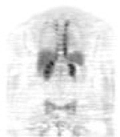
 |
| Fifty-four-year-old woman with axillary node-positive infiltrating lobular carcinoma of the right breast. Patient had been disease-free on systemic hormonal therapy until six years after initial diagnosis when tumor markers became elevated. Posterior coronal image from FDG-PET shows no increased uptake at corresponding skeletal sites. Activity in paraspinal regions is due to muscular uptake. Because of newly diagnosed skeletal recurrence, patient was started on systemic chemotherapy. Eubank W, Mankoff D, Bhattacharya M, Gralow J, Linden H, Ellis G, Lindsley S, Austin-Seymour M, Livingston R, "Impact of FDG PET on Defining the Extent of Disease and on the Treatment of Patients with Recurrent or Metastatic Breast Cancer," (AJR 2004; 183: 479-486). |
"FDG-PET altered the management plan most frequently in patients suspected of having locoregional disease," Eubank said. "The overall sensitivity, specificity, and accuracy of FDG-PET was 94%, 91%, and 92%, respectively."
FDG-PET also proved useful in patients with disease recurrence, suggesting that findings can increase an oncologist's confidence in implementing a specific treatment plan. Eubank's group cited an example of two patients whose treatment was withheld based on PET results. In both cases, the patients avoided the experience and cost of unnecessary treatment.
The group concluded that about one-third of their advanced breast cancer patients were referred for PET imaging to evaluate response to therapy. "This referral pattern underscores the difficulty in evaluation response to treatment with (conventional imaging) in many patients with advanced breast cancer," they said.
By Shalmali PalAuntMinnie.com staff writer
August 16, 2003
Related Reading
NSECT may trump traditional imaging by spotting breast cancer before it manifests, July 28, 2004
PET Medicare reimbursement -- narrow but widening, June 17, 2004
15O-water PET shows persistence of blood flow in chemo-resistant breast tumors, November 28, 2003
Copyright © 2004 AuntMinnie.com


