Hands down, CT angiography beats DSA for aneurysmal treatment planning, said two presenters at the 2001 European Congress of Radiology in Vienna. Where the latter sometimes leaves critical questions unanswered, CTA can reliably depict the morphology of aneurysms and their relation to surrounding structures. That is, except when DSA does a better job.
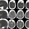
"Digital subtraction angiography [DSA] is the standard imaging modality for visualizing cerebral intracranial aneurysms because of its excellent temporal visualization," said Dr. Csilla Balassy, a radiologist from the University of Vienna. "DSA, however, provides two-dimensional images, and cannot show bone structures very well, so in the case of intracerebral aneurysms, it doesn't always provide all the necessary information for therapy planning."
Because CTA with multiplanar image processing does enable 3-D visualization of intracranial vasculature, the researchers sought to assess its value in depicting complex aneurysms and their relation to surrounding structures, thus providing more clinically relevant information for surgical planning.
The study looked at 19 patients, all of whom had undergone DSA, which did not provide sufficient information for surgical planning. As a result, their aneurysms were deemed complex intracranial aneurysms, Balassy said.
In the study, the treating neurosurgeon developed specific treatment questions for each patient based on DSA results. Then each subject underwent multislice CTA in the specific region of interest.
Images were acquired on a Somatom Plus 4 Volume Zoom scanner (Siemens, Erlangen, Germany) with detector collimation of 2 x 0.5 mm, table increment of 1 to 1.2 mm, slice thickness of 0.5- 0.75 mm, and section spacing of 0.3 mm. Intravenous contrast administration of 60-180 ml was administered to each patient. The resulting images were reconstructed using Vitrea software (Vital Images, Minneapolis).
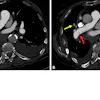
Two radiologists examined the postprocessed images in an effort to visualize the anatomy and answer the specific treatment questions. Following their analysis, 8 patients underwent surgery for aneurysmal repair. The radiologic findings were then compared to surgical findings in these patients, and the therapeutic impact of CTA was evaluated for each of these patients based on a prospective screening, Balassy said.
 |
In 2 patients, complex aneurysms were found at surgery that were not previously demonstrated on CTA images. CTA ruled out one aneurysm, and in 5 patients the CTA was essential for making the decision for or against aneurysmal therapy, Balassy said.
However, Balassy said there are limits to CTA's effectiveness, and cases where DSA can't be replaced. For example, patients who have undergone previous aneurysmal therapy often have aneurysmal clips in the region of interest. These create artifacts on CTA images that can inhibit visualization, she said, especially when the treating surgeon needs information about the morphology of the clipped aneurysm.
With CTA, the presence of small vessels, usually veins, near the region of interest also complicates the evaluation of perforating arteries from the aneurysmal sac, and it is sometimes difficult to determine the origin of particular vessels, she added.
"If we change the window settings [to] get sharper vessel contours, small vessels may disappear. If the setting is so that we see more of the smaller vessels, the [vessel] contours will be more and more [ragged]. It's not easy," Balassy later wrote in an e-mail to AuntMinnie.com. "Because the resolution of DSA is better than CTA, very small aneurysms or perforating arteries may not be detected with 100% sensitivity."
Concluding her presentation, Balassy said that despite its limitations, CTA almost always answers relevant treatment questions.
"In only one patient was CTA not helpful, due to metal artifacts, so altogether 18 of our patients had great impact from CTA, and only one patient was not helped by CTA. We can conclude that CTA provides more therapeutically relevant information than DSA, especially in patients who have giant aneurysms, aneurysms located in the posterior fossa, or (when determining) the exact relation of the aneurysm with the small vessels or bone structures is necessary."
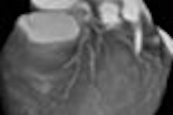
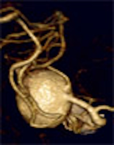
A second presentation that morning reached similar conclusions. In the study, Dr. Zoran Milosevic from the Clinical Institute of Radiology in Ljubiljana, Slovenia, discussed his group's experience with CTA and MPR (multiplanar reconstruction) of giant aneurysms -- those 25 mm and larger.
"Understanding the exact relationship of aneurysms to the surrounding structures, and [their] morphological properties, are of great value," Milosevic said. CTA provides important information regarding surrounding structures and the aneurysmal neck, as well as the size and configuration of aneurysms and the presence of intra-aneurysmal thrombus.
In the study, 12 patients with aneurysms of 25 mm or larger underwent CTA on a Somatom Plus 4 scanner. Multiplanar 3-D reconstructions were then created from the raw image data.
The researchers evaluated the resulting images to determine the size and configuration of intracranial aneurysms, the presence and extent of intra-aneurysmal thrombus, the aneurysms' relationship to other cerebrovascular or skeletal structures, and the presence and orientation of an aneurysm neck. Results were confirmed with DSA or surgery in all patients.
They concluded that because 3-D reconstructions offer views that are similar to those perceived in normal human vision, CTA provides enhanced understanding of the morphology of aneurysms that is useful in surgical planning.
Finally, an article published in European Radiology found CTA to be very useful in depicting intracranial aneurysms (January 2001, Vol.11:1, pp.123-130).
In a study of 200 patients, Australian investigators found that spiral CTA detected 140 of 144 aneurysms, for an overall sensitivity of 97%, including 30 of 32 aneurysms 3 mm or less in size.
In 38 patients with subarachnoid hemorrhage and negative angiography, CTA found 6 of 7 aneurysms that were ultimately diagnosed. There was no significant artifact in 17 of 23 patients (74%) with aneurysmal clips, and the specificity of CTA was 86%, with 8 false-positive cases, the authors wrote.
By Eric BarnesAuntMinnie.com staff writer
April 20, 2001
Click here to post your comments about this story. Please include the headline of the article in your message.
Copyright © 2001 AuntMinnie.com