Although the use of CT in coronary artery imaging is becoming more widespread, its sensitivity for the detection of atherosclerotic plaque is lower than that of traditional invasive angiography, say German researchers.
Dr. Christopher Herzog and colleagues at the Institute of Diagnostic and Interventional Radiology at Johann Wolfgang Goethe University in Frankfurt, Germany, assessed the value and limits of CT angiocardiography in coronary artery imaging. Herzog discussed his team’s research in a presentation at the 2001 European Congress of Radiology in Vienna.
The researchers set out to determine the benefit of CT angiocardiography in coronary artery imaging by comparing the value of four different CT-based visualization techniques. The group selected 50 patients who underwent both multislice CT as well as invasive angiocardiography. The group studied 29 males and 21 females with a mean age of 54 years and a mean heart rate of 63 beats per minute (bpm).
The CT scans were performed on a Somatom Plus 4 Volume Zoom (Siemens, Erlangen, Germany), collimation of 4 x 1 mm, a pitch of 1.5, and a 500-millisecond rotation time. The authors then used retrospective electrocardiogram (ECG) gating (1.25-mm slice thickness, at a 0.5-mm increment) to reconstruct the images.
Each patient’s CT data set was then examined using four different visualization techniques: axial, multiplanar, 3-D, and virtual endoscopy. The data set visualization was conducted on four independent workstations. The axial images were reconstructed on a Somatom Plus 4 Volume Zoom workstation using a 512 x 512 matrix and a slice thickness of 1.25 mm.
 |
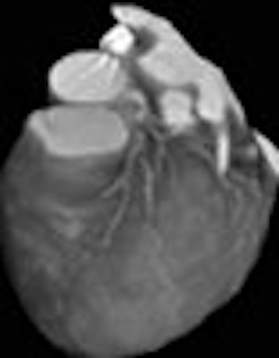
The researchers used a Virtuoso V20A (Siemens) workstation to create the 3-D images, and employed a 256 x 256-pixel matrix in the reconstructions. The virtual endoscopy and multiplanar images were composed using the same application on different machines, but using 256 x 256 and 512 x 512 matrices, respectively. Two radiologists and one cardiologist then evaluated the images for stenoses.
 |
The highest sensitivity for the evaluation of stenoses, 66.7%, was achieved by the axial reconstructions, Herzog said. Virtual endoscopy had a sensitivity of 55.9%, multiplanar reconstructions, 46.6%, and 3-D reconstructions, 33.3%.
However, the techniques offered comparable sensitivities in the detection of atheromatous plaques: 71.2% for the axial scans, 70.1% for 3-D reconstructions, and 69.1% for virtual endoscopy. The multiplanar reconstructions showed distinctly lower results (55.6%) in this test.
The researchers observed that by combining the four techniques, a sensitivity of 74.2% could be achieved. Moreover, the specificity of all four visualization techniques was 91.9%. Herzog said the best CT angiocardiography results were observed at heart rates below 60 bpm.
"Our research showed that CT angiocardiography is restricted to the main branches of the coronary arteries, and that, in the detection of atherosclerotic plaques, it does not achieve a [sensitivity] comparable to invasive angiocardiography," said Herzog.
By Jonathan S. BatchelorAuntMinnie.com staff writer
April 19, 2001
Related Reading
New CT ventriculography technique shows promise, April 13, 2001
MR contrast poised to move forward with USPIO, March 16, 2001
CT angiography best for proximal sequences, lower heart rates, larger vessels, November 29, 2000
Click here to post your comments about this story. Please include the headline of the article in your message.
Copyright © 2001 AuntMinnie.com