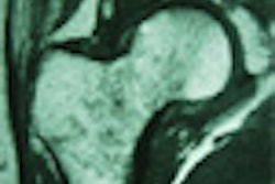
CT ventriculography has gained widespread acceptance as a noninvasive testing method for heart disease. However, the common use of electrocardiogram (ECG) gating in performing the test results in a high radiation dose to the patient due to overlap scanning. A group of Japanese radiologists believe they have found a way to conduct CT ventriculography without the need of an ECG-gating apparatus.
Dr. Seiki Hamada from the Osaka Graduate School of Medicine presented the results of his group's research at last month's European Congress of Radiology in Vienna. The team investigated a new method of cardiac helical multidetector-row CT (MDCT) without ECG gating. To assess the validity of their method, they also conducted retrospective ECG-gated subsecond helical MDCT scans.
The technique utilizes 0.5-mm slice thickness with an acquisition time of 0.5 seconds on a helical MDCT. With a single breath-hold, the patient's entire heart was scanned without ECG gating, at a 5.5-pitch scan, on an Aquilion MDCT scanner (Toshiba, Tokyo). The researchers then reconstructed isotropic slices covering all cardiac phases periodically on a workstation with Plug n View 3D software (Voxar, Edinburgh, Scotland).
Hamada’s group used a four-step method to conduct the ventriculography test on a cardiac motion phantom with contrast media. First they created a coronal multiplanar reformatted (MPR) image from the slices. Then, from the MPR image they re-extracted slices of convex- (top) level as end-diastole, and of concave- (bottom) level as end-systole.
 |
The third step was to create a 3-D reconstruction of end-diastole slices and end-systole slices using interpolation. Finally, the group calculated the phantom volume by measuring the stationary state of the diastolic and systolic phases using conventional helical scanning. The results were compared to ECG-gated MDCT scans of the phantom, and a 1% difference in volumetry was observed.
Based on the success of the cardiac motion phantom, the team applied its technique to the evaluation of the cardiac function in 20 patients. Then retrospective ECG-gated MDCT imaging was performed on another 20 patients, and both methods were compared with conventional ventriculographic methodology.
 |
Hamada reported close correlation between his team’s new technique and the conventional left ventriculographic method (r=0.99, 0.98, and 0.91, for end-diastolic volume, end-systolic volume, and left ventricular ejection fraction, respectively; p<0.05 for all values). He also noted that there was no significant difference between the non-ECG-gated and retrospective ECG-gated MDCT method. The greatest benefit from the new procedure was the low radiation dose on cardiac function analysis due to the high table translation, Hamada said.
By Jonathan S. BatchelorAuntMinnie.com staff writer
April 13, 2001
Related Reading
CT angiography best for proximal sequences, lower heart rates, larger vessels, November 29, 2000
Click here to post your comments about this story. Please include the headline of the article in your message.
Copyright © 2001 AuntMinnie.com