Neurology 1993 Oct;43(10):1966-80
Frontal lobe epilepsy: clinical seizure characteristics and localization with
ictal 99mTc-HMPAO SPECT.
Harvey AS, Hopkins IJ, Bowe JM, Cook DJ, Shield LK, Berkovic SF.
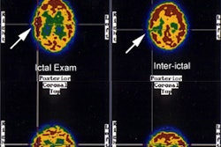
We evaluated ictal 99mtechnetium hexamethyl propylene-amine-oxime single-photon
emission computed tomography (SPECT) in 22 children with electroclinical
features of frontal lobe epilepsy (FLE). Ictal SPECT demonstrated unilateral
frontal hyperperfusion in 20 of 22 children (91%) (one lobar, two frontocentral,
six dorsolateral, six frontopolar, three orbitofrontal, one medial frontal, and
one insula), concordant with electroclinical lateralization in 19 of 20 (95%).
Hyperperfusion was evident in the ipsilateral basal ganglia in 16 of 22 (73%)
and the contralateral cerebellum in 14 of 22 children (64%). Interictal SPECT
showed unilateral, localized frontal hypoperfusion concordant with
electroclinical lateralization in only two of 22 children (9%). Ictal SPECT
localization to the frontocentral, media frontal, or dorsolateral regions was
associated with asymmetric tonic posturing, contralateral head/eye deviation,
and unilateral clonic jerking (p < 0.01). Ictal SPECT localization to the
frontopolar or orbitofrontal regions was associated with vocalization,
hyperventilation, truncal flexion, and complex gestural automatisms (p > or =
0.05). Ictal SPECT has the potential to (1) localize seizures in patients with
intractable FLE, and (2) advance understanding of the in vivo anatomico-clinical
relationships of frontal lobe seizures.