Because some women undergoing uterine fibroid embolization (UFE) may also require ovarian artery embolization (OAE), aortography is routinely performed to assess ovarian arterial supply. But eliminating this step, especially because less than 1% of UFE patients have significant collateral artery supply, could cut procedure-related radiation dose by 20%, according to a Washington, DC-based group.
Medical student Amy White and colleagues from Georgetown University conducted a prospective study on 25 patients undergoing UFE. "Although several investigators have evaluated the patient radiation exposure associated with UFE, none have determined the specific contribution of aortography to the overall radiation exposure," explained White and her co-authors, Dr. Filip Banovac and Dr. James Spies (Journal of Vascular and Interventional Radiology, April 2007, Vol. 19:4, pp. 573-576).
All patients underwent bilateral UFE and one also had right OAE. Embolization was done with either polyvinyl alcohol particles or tris-acryl gelatin microspheres (Contour, Boston Scientific, Natick, MA; Embosphere, Biosphere Medical, Rockland, MD).
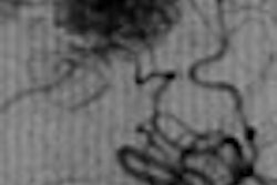
Abdominal aortography was performed in all patients after UFE with images obtained at a rate of one frame every second from the pubic symphysis extending superiorly, the authors said. Angiography was done on a system with an integrated dosimeter (Axiom Artis, Siemens Medical Solutions, Malvern, PA) and gathered data included the dose-area product (DAP).
According to the results, the mean uterine volume was 488.2 cm3 and the mean dominant fibroid volume was 131.2 cm3. In terms of the overall DAP, the main contributor was the embolization process itself, contributing 49% of the total dose. However, 21% of that dose was related to the aortography exam.
"Without routine aortography, we can estimate the average dose reduction to be approximately 20%," the group stated. "We recognize that by not using routine aortography, ovarian vessels providing supply to the uterus will not be detected in a small number of patients. However, it is not clear in every case that the ovarian flow that is detected is supplying fibroids."
The authors conceded that the small number of patients was a limitation to their study and that radiation dose may differ depending on the individual interventionalist's technique. However, they asserted that the study results should encourage clinicians to reconsider routine aortography during UFE.
Instead, based on clinical factors and angiographic findings, it would be more prudent to identify the subset of patients who would benefit from post-UFE aortography, they suggested. In addition, reducing dose during angiography is possible based on their protocol of a single frame per second and no preliminary iliac arteriography.
By Shalmali Pal
AuntMinnie.com staff writer
May 18, 2007
Related Reading
ISET talks stress imaging know-how in AAA endograft, relative safety of UFE, February 12, 2007
Uterine artery embolization for fibroids speeds recovery, January 25, 2007
Tumor regrowth generally calls for second UAE procedure, January 23, 2007
UAE with PVA microspheres yields sustained symptom improvement, September 18, 2006
Copyright © 2007 AuntMinnie.com