When your practice includes a variety of orthopedic exams such as weight-bearing, scoliosis, and long-leg imaging, the current digital radiography offerings may not be versatile enough. At least that's what one large group found when it started going digital, but ended up with computed radiography.
The Illinois Bone and Joint Institute, an 80-physician practice of orthopedists and rheumatologists with several offices in the Chicago area, originally aimed to switch from conventional imaging to a mixture of DR and CR. But the group wound up removing its DR setup and replacing it with another CR room due to various issues, with diminished throughput as the paramount problem.
The institute described its experience with digital conversion in a presentation earlier this year at the American Academy of Orthopaedic Surgeons annual meeting in Washington, DC.
Conversion of three offices to digital imaging began in 2003, as the institute was adding more physicians. Digital radiography was pitched to the group as a way they could meet their increased imaging needs with fewer resources.
"Our thinking was, 'If we did DR, we won't have to build a third x-ray room and hire more staff,'" said Joseph Stark, a registered radiology technologist and PACS manager for the practice.
"That was not the result that we experienced. The DR was much slower than the CR for our needs," Stark said in an interview with AuntMinnie.com.
While the DR was able to process images within seconds, saving a minute or more compared to CR, that time advantage was overwhelmed by the increased time spent in patient positioning, he said.
For example, a three-view study of the knee took seven minutes with the CR machine and 11 minutes with the DR equipment. "That was strictly based on the time that it took to reposition the machine from the AP and lateral to the sunrise view," said Stark.
In addition to spending more time maneuvering the DR's C-arm versus the CR cassettes, the institute's smaller techs often had to double up to roll the DR table around, while the CR had a floating tabletop.
Perhaps most problematic for a practice with many physically impaired patients was the inability to raise or lower the DR system table.
"For weight-bearing foot and ankle exams, we had to have the patient on top of a platform that was higher than this table, and they had to walk up four stairs to get there," Stark noted. "These are patients that are about three weeks post-op. They had a very hard time walking up and down the stairs."
Scoliosis and long-leg exams were simply never performed on the DR system because test runs had shown they would have taken far too long, he said.
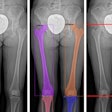
"It would have been about a 15-minute difference for scoliosis exams," said Stark, noting that the physicians required images from the base of the skull to the top third of the femurs, including the greater trochanters. "We would need three images for each view, the AP and the lateral, to cover all that space."
"And then you had to also sit and manually stitch all of those images together," he continued. "We found it to be very inaccurate and very, very time-consuming."
The practice's surgeons also found templating over the DR paper prints to be a problem due to unexpected magnification differences between the DR and conventional radiographs, the institute reported.
Interestingly, the group hadn't anticipated such limitations despite visiting four other orthopedic sites using the DR system. In hindsight, those practices performed more ambulatory orthopedic work or sports medicine that made the DR system a better fit for them, Stark said.
Ultimately, Stark expects that DR will be the way to go for all orthopedic practices. "But it has to be suited to meet the orthopedic needs," he said.
"There's no machine that I've seen that would allow a patient to easily perform weight-bearing lower extremity exams," Stark said. "There's also no machine out there that will do scoliosis and long-leg imaging without spending a gazillion dollars on a 36-inch receptor."
"So there are still a lot of limitations when it comes to the orthopedic aspects of things," Stark said. "Doing the different types of exams that we do day in and day out -- there's such variety in any given day."
By Tracie L. Thompson
AuntMinnie.com staff writer
May 27, 2005
Related Reading
New research shows CR more cost-effective than DR, February 28, 2005
Report finds vendors enable excess radiation doses in pediatric CR, DR, February 4, 2005
Study compares eight digital x-ray systems in clinical setting, December 24, 2004
Computed radiography's (not so surprising) resilience, September 16, 2004
DR looks good in CR, film comparison study, September 19, 2003
Copyright © 2005 AuntMinnie.com

