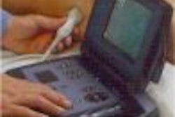
As the use of high-dose fluoroscopy for image-guided procedures increases, so does the potential for radiation-induced injuries to both patients and healthcare workers. This is particularly true when the practitioner has had little to no training in radiation management or on the biological effects of its use.
"In September 1994, the FDA published guidelines for the avoidance of serious x-ray-induced injuries to patients during fluoroscopically guided procedures. Unfortunately, judging by cases recently settled or in litigation, many facilities may be unaware of this advisory document," said Benjamin Archer, Ph.D., a medical physicist at Baylor College of Medicine in Houston.
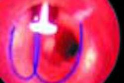
Archer, speaking at the 2001 American Healthcare Radiology Administrators conference in Las Vegas, presented to attendees graphic evidence of high-dose-fluoroscopy-induced dermatitis, epilation, desquamation, and dermal necrosis. The multimillion-dollar settlements that result from these injuries, as well as the potential for increased occupational exposure to physicians and staff, necessitates that an institution have a fluoroscopy-use plan in place, he noted.
Thorough training, competence testing
Because of their familiarity with radiation safety protocols, radiology administrators are the primary people to bring fluoroscopy-use issues to the attention of senior administrators at a facility, Archer said. Another compelling reason to put such a use plan into place is that the Joint Commission on Accreditation of Health Organizations (JCAHO) has taken under consideration the implementation of specific standards and privileges for practitioners to use fluoroscopic x-ray equipment.
Archer, and the American Association of Physicists in Medicine (AAPM), recommend that all practitioners who use fluoroscopy be required to undergo focused training in radiation physics, radiation biology, and radiation safety. This training could include self study, lectures, and hands-on demonstrations with competence testing for physicians seeking fluoroscopic privileges.
This training must be specific to the fluoroscopy systems that the practitioner will be using. Although the same radiological principles apply to all machines, each manufacturer’s system will have features and controls that are unique to it. Archer recommended that the vendor’s application specialists conduct staff instruction on any new equipment brought into a facility, as well as periodic review sessions on existing fluoroscopy installations.
Protocol and technique
Archer listed several factors that influence patient skin dose during fluoroscopy procedures. Along with proper technique that accounts for the size of the patient, positioning of the equipment relative to the patient is crucial.
"I’ve actually seen situations where nonradiologist physicians have confused the x-ray tube for the image intensifier, placing the tube close to the patient. In a couple of reported cases, the spacing device was actually removed from the tube and the tube was placed directly in contact with the patient’s skin," Archer said.
Other dose-reduction factors that were noted include careful use of image magnification by using the least possible magnification for a procedure. Grid removal should also be considered when a high-dose procedure is scheduled. This is especially true for pediatric cases or small adult patients.
Tightly collimating the area of interest for the procedure will reduce scatter radiation, resulting in lower exposed tissue volume for the patient and lower dose rates for personnel performing the exam. Beam-on time, however, is the single greatest factor that can be controlled in fluoroscopy.
"I’ve observed practitioners who work the cine pedal like it was the accelerator pedal in a car," observed Archer.
Because recording images typically involves higher dose rates, image acquisition should be limited to only those captures necessary for the procedure. Archer recommends a policy of always using the five-minute timer, and logging cumulative exposure times for each procedure. In addition, the last-image-hold feature -- if available -- should be exploited as much as possible, as it will allow the physician time to view the exam's progress without exposing the patient to additional radiation.
QC and monitoring
Although training, technique, and protocol are of paramount importance, they can only mitigate the potential damage from an improperly calibrated system. If a system has degraded in its performance characteristics, some operators may be tempted to increase dose rates to compensate. Therefore, periodic evaluations performed by a qualified medical physicist should include an image quality evaluation, as well as documented compliance with state and U.S. regulatory requirements, Archer said.
A facility also may want to consider implementing a patient-dose monitoring system for its high-dose fluoroscopy unit. According to Archer, there are several devices available on the market that will monitor patient dose -- such as thermoluminescent dosimeters, scintillation dosimeters, dose-area product meters, and computerized dose monitors -- which will not interfere with the performance of the interventional procedure.
While the ultimate responsibility for patient safety lies with the physician performing the interventional procedure, hospitals and administrators should proactively require training and certification in radiation management, Archer said. The possibility of litigation should not be the primary motivator for creating a fluoroscopy-use plan.
"Even one radiation injury caused by lack of education is unacceptable," he said.
By Jonathan S. BatchelorAuntMinnie.com staff writer
October 11, 2001
Related Reading
CT dose study is good news for interventional radiologists, October 5, 2001
Low-dose CT fluoroscopy minimizes interventional exposure, July 30, 2001