The first results of the Digital Mammographic Imaging Screening Trial (DMIST) trial may be in, but breast imaging specialists aren't resting on their laurels. While a group from Texas forges ahead on improving full-field digital mammography (FFDM) systems from a technical standpoint, the RSNA is looking to help readers brush up on their interpretive skills using this new technology.
Mo' better filter
According to the DMIST results, FFDM proved better than film-screen (FSM) in women with dense breasts, but was not markedly better in the general population as a screening tool. One possible reason for this outcome may lie in the way FFDM systems are constructed. Investigators from the University of Texas Medical Branch in Galveston have proposed that the standard thickness of a molybdenum (Mo) filter used in FFDM is not optimal. In fact, they found that twice the filter thickness of standard FFDM systems was more appropriate.
"Currently, all digital mammography units still incorporate the same x-ray tube filtration (0.03-mm Mo and 0.025-mm rhodium [Rh]) inherited from their film-screen predecessors," wrote Dr. Thomas Nishino and colleagues. "This level of x-ray tube filtration, although roughly adequate for film-screen mammography systems, may not be optimal for digital mammography" (American Journal of Roentgenology, October 2005, Vol. 185:4, pp. 960-963).
Basic differences in FFDM and film-screen systems include disparate detectors as well as varying responses to x-ray spectra and fluence, they added. Nishino's group conducted phantom experiments on an amorphous silicon-based FFDM detector (Senographe 2000D, GE Healthcare, Chalfont St. Giles, U.K.). They chose the GE system because it is the most widely used FFDM system on the market, they said.
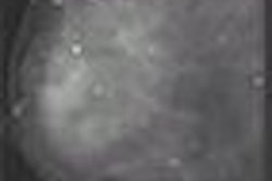
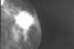
They compared Mo filters of different thicknesses while imaging a custom-made 5-cm thick breast phantom composed of 50% adipose tissue and 50% glandular tissue. Two lesion-simulating inserts were place in the phantom.
The x-ray tube was set to 27 kVp and 200 mAs. Custom-made Mo filters that ranged from 30-90 µm in thickness were inserted into the filter wheel in increments of 15 µm for phantom image acquisitions. The authors compared the lesion-tissue contrast-to-noise ratio (CNR) and radiation dose to the breast.
"The filter shapes the incident x-ray spectrum and therefore greatly affects the image CNR and radiation dose," they explained.
According to the results, the optimal Mo filter thickness in FFDM was 60 µm rather than 30 µm. In their breast phantom, the squared lesion-tissue CNR per unit dose increased with the Mo filter thickness and reached a plateau from 75-90 µm. The increase in filter thickness resulted in a minimal increase in exposure time (three seconds), the authors stated. Finally, this improvement in CNR per dose can be achieved without any increase in the cost of FFDM equipment.
The authors acknowledged certain limitations to this experiment, such as its one-size-fits-all approach to breast thickness, simulated lesion insert, and peak kVp. However, they said they believe that 30 um is a subpar filtration thickness in FFDM.
"We hope that our results will stimulate more work in filtration optimization research from the radiology community and the imaging equipment industry," they wrote.
In an e-mail interview with AuntMinnie.com, Nishino stated that a change in filter thickness would not have made a tremendous difference in the outcome of the DMIST trial. He stressed that "our results of improved CNR per unit dose were only tested under very specific conditions. A broader experiment would be required."
Nishino wrote that GE and other FFDM vendors are aware of his group's findings, which were presented at the 2005 American Association of Physicists in Medicine (AAPM) meeting in Seattle, and have "shown interest in the results. However, we are unaware of any filter thickness changes made by any major vendors."
"Our results are valid with any system that utilizes the same type of detector used by the Senographe 2000D," Nishino added. "Slight modifications in the simulations would be necessary to make the results viable with other detectors (e.g., amorphous selenium). Regardless of the detector, as long as it is a digital ... current filter thicknesses for both Mo and Rh are not optimal for FFDM."
Fine-tuning FFDM skills
In other FFDM news, the RSNA will offer an FFDM self-assessment workshop at the 2005 meeting in Chicago. These interactive seminars will start on November 27 and run through December 1. Each session will last for one hour, with a maximum of 24 participants at a dozen workstations. Eight datasets will be available for review, with participants receiving immediate feedback. CME credit is available for each completed dataset, and the workshop should meet the physician self-assessment modules (SAMs) certification by the American Board of Radiology.
After the seminar, participants can shift to a discussion area to talk with one of the following workshop faculty members:
- Dr. Ulrich Bick from the University Clinic Charité in Berlin
- Dr. J. Timothy Blackwelder from Loma Linda University School of Medicine in Loma Linda, CA
- James Culley, Ph.D., from Hologic in Bedford, MA
- Dr. Carl J.G. Evertsz from MeVis BreastCare in Bremen, Germany
- Dr. Roland Holland from the National Expert and Training Center for Breast Cancer Screening, in Nijmegen, Netherlands
The workshops will be held in Room E266 in the Lakeside Center at McCormick Place. There is no additional fee to attend a workshop, but preregistration is strongly recommended at rsna2005.rsna.org.
By Shalmali Pal
AuntMinnie.com staff writer
October 3, 2005
Related Reading
DMIST study: Younger women may benefit most from digital mammo, September 16, 2005
Need SAMs for recertification? ARRS offerings prove popular, June 1, 2005
Anode/filter combination enables lower dose in FFDM, March 6, 2005
Copyright © 2005 AuntMinnie.com