Adenoid Cystic Carcinoma:
Clinical:
Adenoid cystic carcinoma is a low grade malignancy that arises from epithelial glands lining the respiratory tract- it is a salivary gland type carcinoma. It is the second most common tracheal tumor after squamous cell carcinoma and it is the most common salivary gland type carcinoma of the lung (accounting for 75% of reported cases), followed by mucoepidermoid carcinoma (5-10% of cases) [2]. The lesion is usually found in the lower trachea or mainstem bronchi (90%)- a peripheral or segmental location is uncommon [4]. It is the second most common malignancy of the trachea (accounts for 25-33% of tracheal tumors- squamous cell carcinoma is the most common tracheal tumor) [1,2]. Patients are usually in their 40's-50's (nearly half of affected patients are younger than 30 years of age [3]) and may present with symptoms of obstruction such as wheezing, cough, or dyspnea. There is no association with smoking. The tumor has an equal sex distribution [4].Because of its submucosal origin, the lesion usually grows along the tracheobronchial walls infiltrating the submucosa over long segments, is locally invasive, and frequently recurs locally after resection (the longitudinal extent is typically greater than the cross-sectional area [3]). Metastases to regional lymph nodes are seen in up to 10% of patients [2].
X-ray:
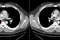
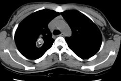
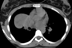
On CT the lesion appears as an endobronchial mass which constricts the tracheal or main bronchial lumen due to submucosal growth which circumferentially encases the airway [6]. The lesion can also produce a circumferential thickening of the tracheal wall, or appear as a soft tissue mass filling the airway [1,2].Lesions located in the lung periphery (about 10% of cases) appear as a solitary pulmonary nodule. FDG uptake is commonly seen with high and intermediate grade tumors [2].
REFERENCES:
(1) J Thorac Imag 1995, 10: p.180-198
(2) AJR 2004; Kwak SH, et al. Adenoid cystic carcinoma of the airways: helical C and histopathologic correlation. 183: 277-281
(3) AJR 2007; Jeong SY, et al. Integrated PET/CT of salivary gland type carcinoma of the lung in 12 patients. 189: 1407-1413
(4) Radiographics 2009; Park CM, et al. Tumors in the
tracheobronchial tree: CT and FDG PET features. 29: 55-71
(5) AJR 2013; Ngo VH, et al. Tumors and tumorlike conditions of
the large airways. 201: 301-313
(6) Radiographics 2018; Lichtenberger JP, et al. Primary lung tumors in children: radiologic-pathologic correlation. 38: 2151-2172


