Pulmonary Arteriovenous Malformation
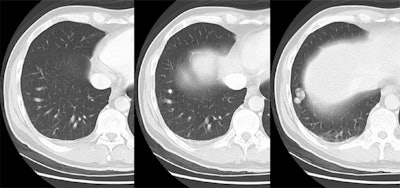
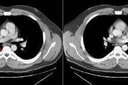
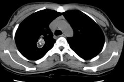
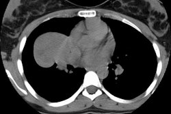
The patient shown below presented with a brain abscess. Portable plain films were of poor quality so a chest CT was done to assess for the presence of a pulmonary AVM. The lesion can be seen in the right costophrenic sulcus with a single artery and a single draining vein (simple AVM).
Cases are best viewed with a cine format which will aid in identification of the arterial and venous communications. Click here to view cine images.


